SHARE Knowledge Hub
Ionising Radiation Diseases
Ionising radiation can cause acute tissue injury after high doses and can increase long-term cancer risk at lower or prolonged doses. Acute radiation syndrome requires a high, penetrating, short-duration exposure to much of the body; local skin injury and internal contamination follow different patterns. Workers should report abnormal exposure promptly so the source can be controlled, dose reconstructed and specialist medical and radiation advice arranged.
Understanding Ionising Radiation Diseases
Ionising radiation can cause acute tissue injury after high doses and can increase long-term cancer risk at lower or prolonged doses. Acute radiation syndrome requires a high, penetrating, short-duration exposure to much of the body; local skin injury and internal contamination follow different patterns. Workers should report abnormal exposure promptly so the source can be controlled, dose reconstructed and specialist medical and radiation advice arranged.
Why this matters at work
Stochastic effects are changes in probability, principally cancer risk, rather than a predictable symptom in each exposed individual. A low dose usually causes no observable immediate illness, but work exposure should still be restricted. A later cancer cannot normally be attributed to one exposure from symptoms alone; an occupational attribution requires clinical, exposure and epidemiological evidence.
An uncontrolled high exposure may cause skin redness or burns after a delay, eye injury, nausea, weakness or other symptoms depending on the exposure. These symptoms are non-specific: heat, chemical exposure and illness can look similar. A person who feels well after a suspected radiological event still needs an exposure assessment. A line manager must not use symptoms to estimate dose or clear a worker to return.
How organisations manage this in practice
Choose controls at source and in design: avoid unnecessary exposure, use enclosures and interlocked shielding, contain unsealed material, provide remote handling and ventilation where appropriate, secure sources and restrict access. Then define local rules, competence, monitoring and supervision. Plan safe stop and recovery if an interlock fails or a source is not returned to the shielded position.
Review after a source, layout, task, worker group or monitoring result changes, and after incidents. Test whether controls work under actual operating and maintenance conditions. Consult the RPA when required and involve workers who know the task. The risk assessment should drive a verifiable work system rather than merely document a dose estimate.
Audit at the point of work. Test interlocks, observe boundaries during site radiography, sample source inventory and review how contaminated material is handled. Check that dosimeters are worn correctly and readings reviewed, not merely issued. Ask workers and contractors whether local rules are usable at night or during maintenance. Verify corrective actions physically.
What health boundary should readers understand?
This page is workplace guidance, not medical diagnosis or treatment advice.
Managers should not diagnose radiation disease, interpret dose significance alone or reassure workers after suspected overexposure without competent advice. Ionising radiation issues can be technically complex because dose, dose rate, radiation type, body part, contamination route and exposure duration all matter.
If a worker suspects significant exposure, contamination, skin injury, eye exposure, source failure or abnormal dosimetry, the response should involve the radiation protection adviser or relevant competent radiation lead, occupational health or appointed doctor arrangements where applicable, and medical assessment routes where needed.
- Dose result concern — Escalate to radiation protection and occupational health routes. Managers interpreting dose without competence.
- Suspected contamination — Control spread and seek specialist advice. Letting the person leave without assessment where contamination is plausible.
- Skin/eye symptoms — Use medical assessment route and exposure investigation. Assuming symptoms are unrelated.
- Worker anxiety — Communicate facts calmly and confidentially. Minimising concern or sharing dose information casually.
- Long-term risk — Explain prevention and monitoring evidence. Making unsupported statements about cancer risk.
What are the key facts about ionising radiation diseases?
Ionising radiation risk depends on dose, tissue, exposure route and control reliability.
The route matters: external irradiation is different from radioactive material entering the body. Early symptoms alone cannot diagnose dose or disease, and routine low-dose work should not be described as acute radiation syndrome. Dose records, contamination checks and competent interpretation guide follow-up; urgent suspected overexposure warrants the site emergency route and medical advice. See UKHSA basic concepts of radiation; UKHSA CBRN incidents: clinical management and health protection.
Ionising radiation can damage cells
It has enough energy to ionise atoms and can damage DNA or tissue.
Effects can be acute or delayed
High exposures may cause tissue effects; lower exposures are controlled because long-term cancer risk matters.
Contamination is different from exposure
A worker can be irradiated externally or contaminated internally/externally with radioactive material.
Dose records matter
Dose assessment and investigation help show whether exposure controls are working.
Prevention is the main health strategy
Time, distance, shielding and competent controls protect people before disease occurs.
What are ionising radiation diseases in workplace terms?
The phrase covers health effects that can arise from workplace exposure to ionising radiation.
Ionising radiation diseases are not one single illness. The relevant workplace concern may be acute skin injury after a high local exposure, cataract risk to the lens of the eye, contamination and internal dose, blood effects after significant exposure, or increased cancer risk later in life.
In most well-controlled workplaces the objective is to prevent significant dose and detect any unusual exposure early. That means a disease page still has to focus heavily on management controls: source inventory, competent advice, local rules, dose monitoring, training and emergency response.
- External exposure — Radiation reaches the body from a source outside it. X-ray set, radiography source or sealed gauge.
- Contamination — Radioactive material is on or in the body or workplace. Unsealed source work, spills or dust/aerosol contamination.
- Absorbed/effective dose — Ways of describing energy deposited and health relevance. Dose records and dose restriction.
- Deterministic/tissue effects — Effects with thresholds where severity increases with dose. Skin burns, cataract and acute injury concerns.
- Stochastic effects — Probability of effect increases with dose, with no simple threshold. Long-term cancer-risk prevention.
How can ionising radiation harm the body?
Ionising radiation can directly or indirectly damage cells and DNA.
The biological mechanism matters because it explains why exposure is controlled so carefully even where workers feel well. Ionising radiation can break chemical bonds, generate reactive molecules and damage DNA. Some damage is repaired by the body; some may lead to cell death; some may increase the probability of later disease. See UKHSA basic concepts of radiation.
This is why radiation protection is preventive and evidence-led. Waiting for symptoms would be the wrong strategy. Dose restriction, monitoring and investigation levels exist to prevent harm before obvious illness appears.
- DNA damage — Long-term cancer risk. Keep dose ALARP and monitor exposure.
- Cell killing at high dose — Tissue injury or acute effects. Prevent high-dose exposure and control access.
- Lens exposure — Cataract risk. Eye dose control and shielding where relevant.
- Internal contamination — Dose to organs from inhaled/ingested material. Containment, hygiene and contamination monitoring.
- Local skin dose — Skin injury from close or direct exposure. Shielding, distance and source handling controls.
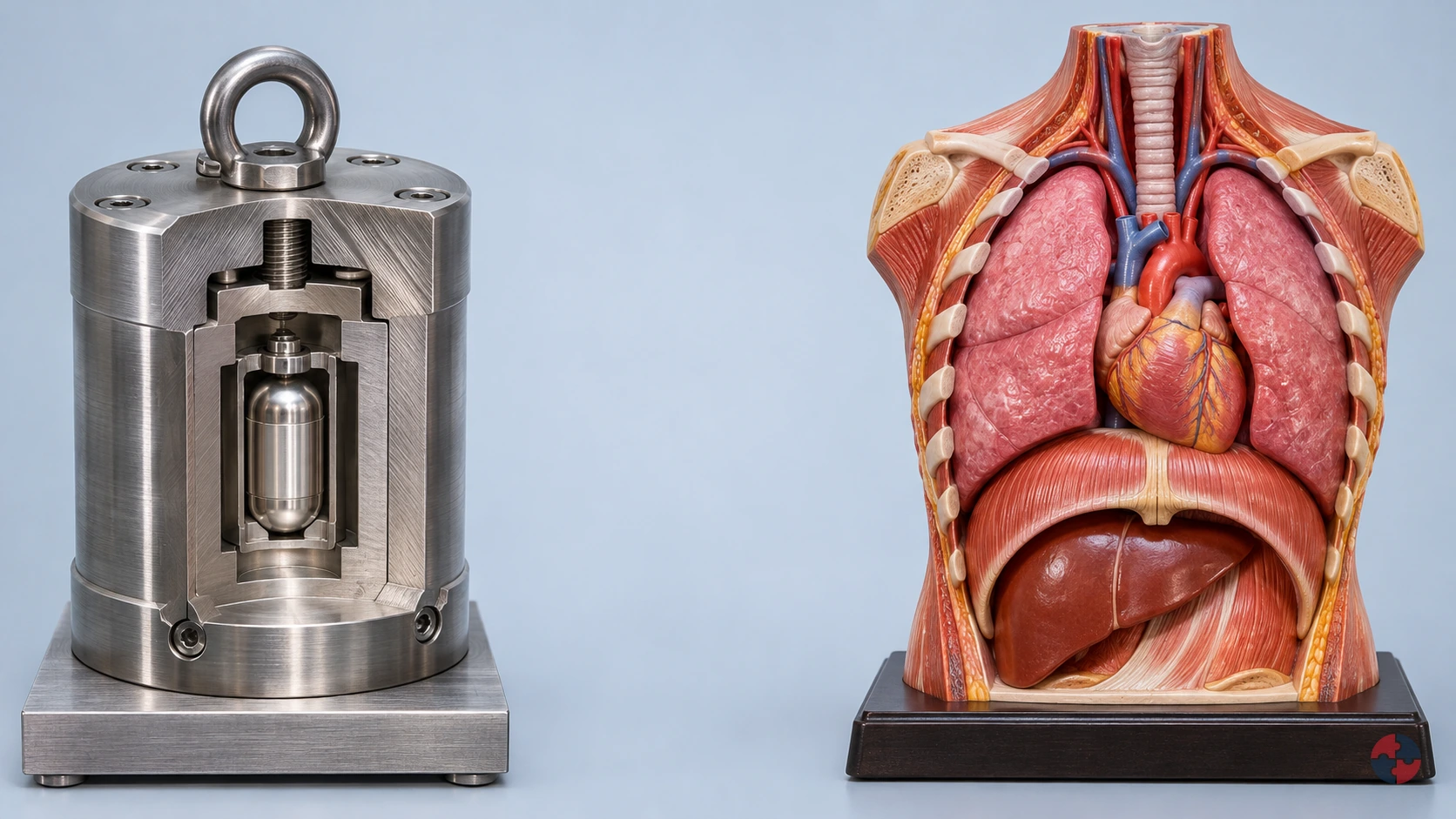

How can different types of ionising radiation reach and damage body tissues?
Interactive radiation explainer · SHAREhub environment
Read all five radiation, shielding and body-effect explanations
Alpha
Typical sources: Alpha-emitting material Real example: radon and its radioactive decay products in buildings or underground workplaces. An unstable nucleus emits an alpha particle: two protons and two neutrons. The display holder is a teaching cutaway, not a radon container or an equipment design.
Travel and distance: A few centimetres in air Typical alpha particles rapidly lose energy in air. The illustrated track length is enlarged.
Penetration and exposure route: External: stops at the outer dead skin layer This applies to intact outer skin. Inside an airway or a wound, living cells have no such protective barrier.
Possible effects: A short, dense ionisation track can damage nearby cells. Inhaled radon decay products expose lung tissue and increase lung-cancer risk.
When radioactive material enters the body: Internal alpha: an airway deposit Illustrates an inhaled radioactive decay product near airway cells. Its short range concentrates energy locally; the exact organ dose depends on the radionuclide and where material is retained. 1 · A radioactive deposit is already inside The airway inset starts after an alpha-emitting radioactive decay product has been inhaled and deposited beside living cells. It can continue to emit radiation while it remains there. 2 · Follow a short track through local tissue The particle now starts inside the airway lining, rather than outside intact skin. It deposits energy densely over a short local track; short range does not make an internal alpha emitter harmless. 3 · Why an outside barrier cannot shield this deposit Paper outside the body cannot sit between the deposited emitter and adjacent cells. Containment and preventing intake address the exposure route; an exterior shield does not remove material already inside. 4 · Examine local cell interactions Nearby cells may receive energy that damages biological molecules, including DNA. Radon-related exposure can increase lung-cancer risk over time. The highlighted cells do not predict an immediate injury, a measured dose or a certain cancer outcome.
Shielding and control choices: Paper illustrates alpha stopping; preventing radioactive material entering the body is the crucial additional control. A paper barrier does not control airborne radon or contaminated dust.
Example sequence: 1 · See the source The teaching holder reveals alpha emissions. Radon-related exposure usually comes from radioactive gas and its decay products, not a visible glowing rock. 2 · Follow the short air track The particle deposits energy densely and stops over a short path. A few centimetres is a useful typical scale, not a clearance distance. 3 · Insert a barrier A sheet of paper stops the illustrated alpha track. The source remains radioactive even when no track reaches the body. 4 · Compare outside and inside Outside: the track stops at intact outer skin. Select internal exposure to see an airway deposit irradiate adjacent living cells. This body inset is a separate unshielded local comparison; it does not show the outcome of the selected barrier.
Compare barrier materials
- Air only: The short alpha track stops in air before distant tissue; close or internal exposure is a different situation. Material comparison only; thickness, energy and geometry are not calculated.
- Paper: Paper stops the illustrated alpha particles. Containment and control of intake still matter. Material comparison only; thickness, energy and geometry are not calculated.
- Plastic: Plastic stops the illustrated alpha particles. Containment and control of intake still matter. Material comparison only; thickness, energy and geometry are not calculated.
- Aluminium: Aluminium stops the illustrated alpha particles. Containment and control of intake still matter. Material comparison only; thickness, energy and geometry are not calculated.
- Lead: Lead stops the illustrated alpha particles. Containment and control of intake still matter. Material comparison only; thickness, energy and geometry are not calculated.
- Concrete: Concrete stops the illustrated alpha particles. Containment and control of intake still matter. Material comparison only; thickness, energy and geometry are not calculated.
- Water: Water stops the illustrated alpha particles. Containment and control of intake still matter. Material comparison only; thickness, energy and geometry are not calculated.
- Layered neutron barrier: Layered neutron barrier stops the illustrated alpha particles. Containment and control of intake still matter. Material comparison only; thickness, energy and geometry are not calculated.
Beta
Typical sources: Beta-emitting material Examples include radioactive tracers in laboratories and some industrial thickness gauges. This example shows beta-minus radiation: an electron emitted during nuclear decay. Generic teaching holder; real sources have purpose-designed containment and protection.
Travel and distance: Centimetres to metres, depending on energy Beta emitters have different energy distributions. Some beta particles travel several metres in air; low-energy examples travel much less.
Penetration and exposure route: Skin and superficial tissue; depth varies More energetic beta particles can reach living skin and the lens of the eye. Do not treat beta as harmless because it is less penetrating than gamma.
Possible effects: External beta exposure can injure skin or eyes at sufficient dose. Radioactive material inside the body can irradiate nearby tissue.
When radioactive material enters the body: Internal beta: a local deposit A simplified contaminated-tissue example. The same electron track now starts within the body. Actual distribution depends on the material; inhalation is not the only possible entry route. 1 · A beta emitter is already beside tissue This airway-cell example begins with radioactive material already deposited inside the body. Inhalation is one entry route; other materials can enter by swallowing or through wounds and distribute differently. 2 · Follow an electron from within the body The emitted beta electron scatters and loses energy in nearby tissue. Its finite local track depends on energy and tissue; the magnified drawing does not represent a fixed depth or organ dose. 3 · Separate outside shielding from internal exposure An external plastic panel cannot be placed between this deposited emitter and neighbouring cells. Preventing contamination entering the body matters; changing the outside panel cannot cancel an internal exposure. 4 · See local energy deposition and possible harm Interactions can affect cells close to the radioactive deposit. The amount and distribution of material, its emissions and retention determine exposure; a coloured track does not establish tissue injury or an individual cancer risk.
Shielding and control choices: Low atomic number plastic is a useful first comparison for beta shielding. Slowing beta particles can generate bremsstrahlung X-rays, especially in dense, high atomic number materials. A designed shield may need more than one layer.
Example sequence: 1 · See the beta emitter The electron starts at the nucleus. This is a beta-minus example; positron-emitting sources need additional explanation because annihilation photons are also produced. 2 · Follow the changing track Interactions scatter and slow the electron. Its travel depends strongly on starting energy, so there is no single range shared by all beta sources. 3 · Compare plastic and lead Plastic absorbs the illustrated beta track with less bremsstrahlung than a comparable high atomic number material. Lead is not an automatic first choice. 4 · See superficial energy deposition The enlarged skin section shows a finite electron track in tissue. Internal contamination places the emitter beside living cells; it changes the exposure route. This body inset is a separate unshielded local comparison; it does not show the outcome of the selected barrier.
Compare barrier materials
- Air only: Longer beta tracks may reach exposed skin; range changes with energy. Material comparison only; thickness, energy and geometry are not calculated.
- Paper is not a universal beta barrier: The example passes through paper. Very low energy beta emitters behave differently. Material comparison only; thickness, energy and geometry are not calculated.
- Low atomic number first layer: Plastic illustrates electron stopping while limiting bremsstrahlung production. Material comparison only; thickness, energy and geometry are not calculated.
- Electron stopping with a secondary-radiation check: Aluminium can stop beta particles; energy and shield thickness determine the result. Bremsstrahlung still needs consideration. Material comparison only; thickness, energy and geometry are not calculated.
- Stopping electrons can create X-rays: High atomic number lead can increase bremsstrahlung production. A real beta shield may start with plastic and use another layer for photons. Material comparison only; thickness, energy and geometry are not calculated.
- Concrete: Beta tracks lose energy in material; a substantial barrier can absorb the electrons. Secondary photons and real construction still need assessment. Material comparison only; thickness, energy and geometry are not calculated.
- Water: Beta tracks lose energy in material; a substantial barrier can absorb the electrons. Secondary photons and real construction still need assessment. Material comparison only; thickness, energy and geometry are not calculated.
- Layered barrier: Beta tracks lose energy in material; a substantial barrier can absorb the electrons. Secondary photons and real construction still need assessment. Material comparison only; thickness, energy and geometry are not calculated.
Gamma
Typical sources: Gamma-emitting radioactive source Industrial radiography uses shielded radioactive sources to inspect components such as pipe welds. A nucleus releases a photon as it moves to a lower energy state. Generic capsule in an illustrative cutaway; real radiography sources remain within engineered systems.
Travel and distance: Long-range: attenuates rather than ending at one distance Photons can cross substantial air gaps. Distance changes exposure but does not create a universal safe boundary.
Penetration and exposure route: Can reach deep organs or pass through the body Some photons interact and deposit energy; others pass through. Penetration depends on energy, tissue and geometry.
Possible effects: Energy deposited in tissue can damage cells and DNA. Risk depends on dose and exposed tissue; sufficiently high doses can also cause tissue injury.
Shielding and control choices: Lead and appropriately designed concrete barriers reduce photon transmission. Shielding attenuates the beam. It is misleading to show one standard sheet as stopping every gamma photon.
Example sequence: 1 · See a continuing radioactive emission The capsule emits gamma photons as radioactive decay proceeds. Turning off an electrical supply does not stop radioactive decay. 2 · Follow photons across the scene Unlike the short alpha path, photon transmission falls progressively. The scene is compressed and provides no safe-distance calculation. 3 · Reduce the transmitted beam Absorption and scattering in the barrier leave fewer illustrated forward tracks. Some transmission remains: real design checks also consider scattered radiation and openings. 4 · See interactions at different depths A penetrating photon can transfer energy inside the body. The highlighted points represent interactions, not a prediction of injury or cancer. This body inset is a separate unshielded local comparison; it does not show the outcome of the selected barrier.
Compare barrier materials
- Unshielded path: Photons can continue across the scene and into tissue. Distance and time influence exposure. Material comparison only; thickness, energy and geometry are not calculated.
- Paper gives little protection in this example: The illustrated photon beam largely continues. Paper is not an appropriate photon protection strategy. Material comparison only; thickness, energy and geometry are not calculated.
- Plastic: Some attenuation occurs, but this illustrative layer leaves substantial photon transmission. Energy and actual thickness matter. Material comparison only; thickness, energy and geometry are not calculated.
- Aluminium: Some attenuation occurs, but this illustrative layer leaves substantial photon transmission. Energy and actual thickness matter. Material comparison only; thickness, energy and geometry are not calculated.
- Dense photon barrier: Lead reduces forward transmission through absorption and scattering; some photons still transmit or scatter. Material comparison only; thickness, energy and geometry are not calculated.
- Structural photon shielding: A designed concrete barrier can reduce the field. Composition, thickness, joints and openings affect its performance. Material comparison only; thickness, energy and geometry are not calculated.
- Water also attenuates photons: A sufficient path through water reduces transmission. The small illustrated layer does not establish a protective thickness. Material comparison only; thickness, energy and geometry are not calculated.
- Assess the photon component separately: A neutron-oriented composite is not automatically a complete photon shield. Assess the materials and photon energies actually present. Material comparison only; thickness, energy and geometry are not calculated.
X-rays
Typical sources: Energised X-ray tube Medical and dental imaging, security screening, cabinet inspection and handheld X-ray fluorescence analysis. Electrons accelerated within a tube strike a target and produce X-ray photons. The beam appears only during the illustrated energised exposure. This is not a radioactive source capsule.
Travel and distance: No single stopping distance in air X-rays are photons. Beam energy, filtration, direction and scatter all affect transmission.
Penetration and exposure route: Can penetrate the body; absorption differs between tissues Differences in transmission allow diagnostic images. The examination uses a controlled, justified exposure.
Possible effects: X-rays can damage cells, so exposures are controlled. A normal diagnostic X-ray does not leave radiation stored inside a person or make them radioactive.
Shielding and control choices: Purpose-designed shielding such as lead or concrete reduces the primary and scattered X-ray field. X-rays are not always less penetrating than gamma rays: photon energy, not the name alone, governs their interactions.
Example sequence: 1 · Energise the teaching tube An electron beam meets the target; X-ray photons leave the tube window. A normal diagnostic tube produces its beam while energised. 2 · Follow a directed beam and scatter The illustrated beam travels through air. Some interactions redirect photons; this is why the forward beam alone does not describe every exposure. 3 · Place a designed photon barrier The barrier reduces transmission. Real installations account for equipment output, workload, occupants and scatter, with suitable expert advice. 4 · Compare tissue transmission Some photons cross the body and some interact. The beam ends when the diagnostic exposure ends; the patient does not retain the X-ray beam. This body inset is a separate unshielded local comparison; it does not show the outcome of the selected barrier.
Compare barrier materials
- Unshielded path: Photons can continue across the scene and into tissue. Distance and time influence exposure. Material comparison only; thickness, energy and geometry are not calculated.
- Paper gives little protection in this example: The illustrated photon beam largely continues. Paper is not an appropriate photon protection strategy. Material comparison only; thickness, energy and geometry are not calculated.
- Plastic: Some attenuation occurs, but this illustrative layer leaves substantial photon transmission. Energy and actual thickness matter. Material comparison only; thickness, energy and geometry are not calculated.
- Aluminium: Some attenuation occurs, but this illustrative layer leaves substantial photon transmission. Energy and actual thickness matter. Material comparison only; thickness, energy and geometry are not calculated.
- Dense photon barrier: Lead reduces forward transmission through absorption and scattering; some photons still transmit or scatter. Material comparison only; thickness, energy and geometry are not calculated.
- Structural photon shielding: A designed concrete barrier can reduce the field. Composition, thickness, joints and openings affect its performance. Material comparison only; thickness, energy and geometry are not calculated.
- Water also attenuates photons: A sufficient path through water reduces transmission. The small illustrated layer does not establish a protective thickness. Material comparison only; thickness, energy and geometry are not calculated.
- Assess the photon component separately: A neutron-oriented composite is not automatically a complete photon shield. Assess the materials and photon energies actually present. Material comparison only; thickness, energy and geometry are not calculated.
Neutrons
Typical sources: Neutron-emitting source Examples include specialist research, nuclear facilities and neutron gauges; an americium–beryllium capsule is one neutron-source type. Some high-energy medical accelerators produce secondary neutrons. Nuclear reactions can release uncharged neutrons. This teaching example represents a contained neutron source. Generic source representation; mixed neutron and gamma fields require specialist assessment.
Travel and distance: Energy-dependent; potentially highly penetrating Neutrons scatter and change energy. Neither a fixed air range nor a single lead sheet describes their behaviour.
Penetration and exposure route: Can irradiate deep tissue Neutrons transfer energy through nuclear interactions, producing charged particles that deposit energy locally.
Possible effects: Neutron interactions can damage tissue and increase cancer risk. Biological effect varies with neutron energy; counting tracks alone does not compare doses between radiation types.
Shielding and control choices: Hydrogen-rich material slows fast neutrons; a capture component can remove slowed neutrons. A designed barrier also considers photons. Capture and other interactions can produce secondary gamma radiation. Moderating a neutron is not the same as removing all radiation.
Example sequence: 1 · See an uncharged neutron leave the source The neutron starts from a nuclear process, not from the electron beam of an ordinary diagnostic X-ray tube. Real sources may emit gamma radiation as well. 2 · Follow scattering and changing energy A neutron can travel well beyond the short alpha path. Collisions can deflect it and reduce its energy, rather than simply making it vanish. 3 · Slow, capture and consider secondary photons Hydrogen-rich shielding illustrates moderation, followed by a capture component. A secondary gamma track reminds learners that the whole field must be assessed. 4 · See energy transferred to tissue nuclei An interaction creates an energetic charged recoil or reaction product. The short recoil track represents local energy deposition, not a visible wound or a dose estimate. This body inset is a separate unshielded local comparison; it does not show the outcome of the selected barrier.
Compare barrier materials
- No moderating or capture barrier: Neutrons can reach tissue. Scattering and energy changes make a universal air-range claim unsuitable. Material comparison only; thickness, energy and geometry are not calculated.
- Paper: This is not a complete fast-neutron protection strategy. A material effective for photons may perform poorly for neutrons. Material comparison only; thickness, energy and geometry are not calculated.
- Aluminium: This is not a complete fast-neutron protection strategy. A material effective for photons may perform poorly for neutrons. Material comparison only; thickness, energy and geometry are not calculated.
- Lead: This is not a complete fast-neutron protection strategy. A material effective for photons may perform poorly for neutrons. Material comparison only; thickness, energy and geometry are not calculated.
- Hydrogen-rich plastic moderates: Collisions with hydrogen help slow fast neutrons. Moderation alone does not guarantee capture or control secondary radiation. Material comparison only; thickness, energy and geometry are not calculated.
- Concrete can shield mixed fields: Hydrogen content and composition matter for neutrons; the design must also account for secondary photons. Material comparison only; thickness, energy and geometry are not calculated.
- Water illustrates moderation: Hydrogen helps slow neutrons. Some interactions produce secondary gamma radiation; a real barrier is assessed as a whole. Material comparison only; thickness, energy and geometry are not calculated.
- Moderation plus capture: Hydrogen-rich material slows fast neutrons and a capture component removes some slowed neutrons. Secondary gamma shielding may also be needed. Material comparison only; thickness, energy and geometry are not calculated.
Sources and how to interpret the model
Original teaching geometry and animation. The reference diagram is a layout inspiration; no third-party illustration has been copied. Coloured tracks, counts, timing and materials illustrate mechanisms rather than exposure measurements. This activity is not a radiation protection assessment or a design specification.
- Health and Safety Executive: Ionising radiation: protecting workers and others
- UK Health Security Agency: Basic concepts of radiation
- UK Health Security Agency: Living with radiation
- Health and Safety Executive: Radon in the workplace
- Health and Safety Executive: Introduction to radiation in the workplace
- Health and Safety Executive: Work with ionising radiation that requires registration
- Health and Safety Executive: Industrial radiography – managing radiation risks (IRP1)
- NHS: X-ray
- North Tees and Hartlepool NHS Foundation Trust: My X-ray Examination – What You Need to Know about Radiation and the Risks Involved
- Office for Nuclear Regulation: Shielding and dose rate safety assessment of transport packages (NS-TAST-GD-100)
- Office for Nuclear Regulation: Criticality safety assessment of transport packages (NS-TAST-GD-097)
- Health and Safety Executive: Radiation Protection Advisers
- Health and Safety Executive: Dose monitoring, assessment and recording
- UK Health Security Agency: Accidental neutron exposure in a medical setting: a case study
- Office for Nuclear Regulation: Step 4 assessment of radiological protection for the UK EPR (ONR-GDA-AR-11-025)
How can workers be exposed?
Exposure routes include external irradiation, inhalation, ingestion, skin contamination and wounds.
External exposure can occur from X-ray equipment, sealed sources, gauges and radiography. Internal exposure can occur if radioactive substances are inhaled, swallowed or enter through damaged skin. Radon is an important inhalation example because its decay products contribute to lung dose.
The controls must match the route. Shielding helps external exposure; containment, ventilation, hygiene and contamination monitoring help internal exposure.
- External whole-body exposure — Working near radiography source. Distance, shielding, time and exclusion zones.
- Local external exposure — Hand near source or beam path. Tools, guarding and work method.
- Inhalation — Radon, radioactive dust or aerosol. Ventilation, containment and respiratory control.
- Ingestion — Contaminated hands, food or drink. Hygiene, prohibition of eating/drinking and monitoring.
- Wound uptake — Contamination entering broken skin. Skin protection and wound management.
What health effects should be understood?
Health effects depend on radiation type, dose, dose rate, tissue and individual circumstances.
Prompt tissue injury can follow a high absorbed dose to a local area or much of the body; acute radiation syndrome requires a high, penetrating exposure over a short period to much of the body. Cataracts may develop after a latency and should not be listed as an immediate symptom. Protection records often use equivalent or effective dose in sieverts (Sv); acute tissue-effect assessment uses absorbed dose in grays (Gy), with radiation type, tissue and timing interpreted by specialists. UKHSA basic concepts of radiation; UKHSA CBRN incidents: clinical management and health protection
Managers should understand enough to recognise why controls and records matter, but they should not diagnose. The competent route is radiation protection advice, occupational health, appointed doctor arrangements where applicable, and medical support after suspected significant exposure.
| Health effect | What it means | Workplace learning point |
|---|---|---|
| Skin injury | High local dose may damage skin. | Prevent direct exposure and investigate abnormal source handling. |
| Cataracts | Lens of the eye can be sensitive to radiation exposure. | Control eye dose and use shielding/monitoring where relevant. |
| Blood changes | Significant whole-body exposure can affect blood-forming tissues. | Use medical surveillance and dose investigation routes. |
| Radiation sickness | Acute radiation syndrome follows a high, penetrating, short-duration exposure to much of the body; a high local dose may instead cause local tissue injury. | Emergency planning prevents rare but serious scenarios. |
| Cancer risk | Long-term probability can increase with dose. | Keep exposure ALARP and maintain dose records. |
| Pregnancy and fetal exposure | Prenatal exposure can affect a developing fetus; the outcome depends on dose and stage of pregnancy. This is distinct from an inherited disease claim. | Protect privacy and seek individual radiation-protection and clinical advice before changing duties. UKHSA pregnancy guidance. |
How should acute effects, chronic effects and latency be understood?
A stronger scientific understanding helps learners explain why radiation health controls are preventive, long-term and evidence-led.
Acute effects are associated with higher exposures over a short period or high local exposure to a body part. Examples after serious abnormal exposure can include prompt skin injury, blood effects or systemic radiation illness. Lens injury may become apparent later as a cataract, rather than as an immediate symptom. These are not issues a line manager should try to assess informally; the response must be controlled, technical and medical where needed.
Chronic and delayed effects are different. A worker may have no immediate symptoms, but exposure history still matters because some effects, particularly cancer risk, may have long latency. This is why dose records, investigation levels, classified-person controls and long-term record retention matter. The health record is not bureaucracy; it is the evidence trail that allows exposure to be reviewed years later if needed.
Reproductive and pregnancy-related concerns also need careful handling. The correct approach is not public discussion or assumptions about capability. It is confidential risk review, competent radiation protection advice, occupational health input where appropriate and controls that keep exposure properly restricted. For Great Britain, current HSE dose guidance explains the IRR17 route and the worker’s choice about pregnancy notification; use the separate Northern Ireland regulations for NI duties.
| Topic | What it means | Workplace management implication |
|---|---|---|
| Acute tissue effects | Effects that can occur after significant short-term or local exposure. | Stop exposure, secure the source, obtain radiation protection advice and use medical/OH routes. |
| Skin injury | A local tissue effect that may arise where a source or beam exposes skin. | Use shielding, distance, source handling tools, access control and incident investigation. |
| Eye lens and cataracts | The lens can be sensitive to ionising radiation exposure. | Assess eye exposure, use shielding/positioning and consider monitoring where relevant. |
| Blood and systemic effects | High whole-body dose can affect blood-forming tissues and general health. | Prevent credible high-dose scenarios through engineered control and emergency planning. |
| Cancer latency | Cancer risk may arise years after exposure and may not have early warning symptoms. | Keep dose ALARP, maintain dose records and review unusual exposures. |
| Internal contamination | Radioactive material inside the body can irradiate organs over time. | Control inhalation, ingestion and wound routes through containment, hygiene and monitoring. |
| Reproductive concerns | Individual circumstances may need confidential review and competent advice. | Handle privately and avoid assumptions, discrimination or unnecessary disclosure. |
| Medical surveillance boundary | Health assessment must be performed by competent health professionals where required. | Managers retain work-control records, not confidential medical diagnosis details. |
Why are dose records and exposure history important?
Dose records turn an invisible exposure into evidence that can be reviewed, trended and acted on.
Radiation dose records are not just compliance paperwork. They help identify unusual exposures, assess whether controls are working, support classified-person arrangements and provide long-term evidence if health concerns arise. See HSE - Ionising radiation legal base.
Dose data must be interpreted by competent people. A single number without context can mislead; the work activity, dose type, badge use, body part and investigation level all matter.
- Personal dosimetry — Assesses individual exposure where required. Was the dosemeter worn correctly?
- Area monitoring — Checks exposure around sources and boundaries. Are maps current after equipment changes?
- Investigation levels — Trigger review before limits are approached. Are exceedances investigated?
- Classified-person record — Supports legal and medical surveillance arrangements. Is it retained and controlled appropriately?
- Incident dose assessment — Estimates dose after abnormal event. Was competent radiation advice used?
What are classified persons?
Classified-person arrangements apply where exposure may exceed specified levels under IRR17.
A classified person is not simply someone who works near radiation. Classification is a legal designation based on likely exposure levels. It brings additional requirements around dose assessment, dose records and medical surveillance. See Ionising Radiations Regulations 2017; MS33 - Guidance for appointed doctors on IRR17.
Contractors who occasionally work on sites with ionising radiation sources may also need careful pre-job assessment. The host and contractor need clarity about who controls the source, who assesses exposure and what local rules apply.
- Exposure assessment — Decides whether classification is required. RPA advice and dose estimate.
- Designation — Formal classification by employer where criteria apply. Designation record.
- Dosimetry — Dose is assessed by approved arrangements. Dose record.
- Medical surveillance — Appointed doctor arrangements where required. Health record and fitness outcome.
- Information/training — Worker understands duties and local rules. Training and acknowledgement.
How are ionising radiation diseases prevented?
Prevention relies on minimising exposure and preventing abnormal events.
The hierarchy begins with whether ionising radiation is needed at all. Can the work use ultrasound, visual inspection, non-radioactive measurement, lower activity or better design? Where the source is needed, exposure is restricted through engineering, shielding, containment, distance, time, local rules, controlled areas, dose monitoring and supervision. See L121 - Working with ionising radiation.
PPE is rarely the main answer. The main answer is source control, physical separation, competent planning and disciplined work.
| Control | How it prevents harm | Evidence |
|---|---|---|
| Justify/substitute | Avoids or reduces the radiation source. | Option appraisal. |
| Shield | Reduces external dose. | Shielding survey. |
| Distance and remote handling | Reduces dose rate at the body. | Work method and tools. |
| Limit time | Reduces accumulated dose. | Task plan and rehearsals. |
| Contain contamination | Prevents inhalation/ingestion/skin spread. | Containment and contamination monitoring. |
| Local rules and supervision | Controls behaviour around high-risk sources. | RPS checks and local rules. |
Where might ionising radiation disease risks arise?
Workplace examples help connect disease mechanisms to practical control decisions.
- Industrial radiography — External gamma/X-ray exposure. Exclusion zones, source control, warning systems and trained radiographers.
- Nuclear or research work — External and contamination risks. Containment, monitoring and specialist procedures.
- Scrap/recycling — Orphan source or contaminated material. Detection, quarantine and emergency contact route.
- Manufacturing gauge — Sealed source exposure if damaged or modified. Source inventory, guarding and maintenance control.
- Radon-affected workplace — Inhalation of radon decay products. Measurement, remediation and review.
- Healthcare/veterinary equipment — X-ray exposure during imaging. Shielding, controlled access and authorised operators.
Unexpected dosimeter result after contractor radiography
Task, material and setting: A contractor’s dose badge returns an unexpected high reading after radiography near a fuel terminal. The worker has no symptoms.
People and prerequisites: The site controller excludes exposure and activates the site plan; only trained, specifically authorised responders approach, recover or manipulate a source. The employer, with RPA advice, obtains competent dose and clinical assessment and reconstructs tasks, positions, timing and badge use.
Before: Pause comparable work, preserve the dosimeter and survey records, and follow the employer’s abnormal-dose and emergency procedures.
During: Check whether exposure, contamination or badge misuse is plausible; use competent dose assessment and medical advice as indicated, without diagnosing from symptoms alone.
Hold or stop: Hold radiography if the source, boundary or dose cannot be accounted for; escalate immediately for suspected significant exposure.
Completion and hand back: The radiation and medical specialists set follow-up. Restart only after the exposure pathway and control failure are understood and corrected.
Evidence: Keep source log, permit, local rules, badge and survey data, worker account, specialist advice, reporting decision and control verification.
What medical surveillance and confidentiality issues apply?
Medical surveillance is a defined professional process covering classified employees and people the employer intends to designate as classified, employees who have received an overexposure even if not classified, and specified cases certified by the relevant doctor under the applicable regulations. Classification is not a prerequisite after an overexposure; a supervisor cannot substitute a symptom checklist. The appointed doctor interprets health information within the radiation regime, while the employer retains work-control and dose evidence without distributing private clinical details. See HSE MS33 guidance for appointed doctors.
If a dosimeter alarm follows a source fault, preserve the time, position, device and source information before estimating what happened. The radiation adviser assesses exposure evidence and the appointed doctor handles medical interpretation where the regime requires it; the line manager keeps the worker away from further exposure pending the competent decision. This separates an immediate work-control hold from a diagnosis and prevents sensitive clinical notes being used as an informal permit to return.
- Classified person health record — Maintain required health record and fitness outcomes. Putting detailed medical notes in line-manager files.
- Dose concern — Use appointed doctor/OH and radiation advice. Manager gives informal reassurance.
- Sensitive worker concern — Review risk privately and with competent advice. Publicly discussing pregnancy or health status.
- Incident follow-up — Record exposure facts and health referral route. Speculation about future disease.
- Long-term retention — Retain records as required and control access. Losing dose or health records after role changes.
Use the appointed/relevant doctor route
A non-classified contractor after suspected overexposure still needs the statutory medical-surveillance decision; do not wait for classification. Keep prescribed employer health-record/fitness entries separate from confidential clinical notes. Check GB IRR17 regulation 25 alongside NI regulation 25. The ordinary annual-entry provision has an exception for overexposed non-classified employees; do not apply one universal interval without the relevant doctor/regime decision. See IRR17 regulation 25 in the official instrument.
What should happen after suspected abnormal exposure?
The response should protect people, control the source, preserve facts and involve competent radiation advice.
A qualifying radiation emergency follows the GB or NI emergency regime for the site. Secure the source and seek radiation and medical advice before deciding the reporting and restart route. Radiation (Emergency Preparedness and Public Information) Regulations 2019; Radiation (Emergency Preparedness and Public Information) Regulations (Northern Ireland) 2019; UKHSA CBRN incidents: clinical management and health protection.
- Stop and secure — Prevent further exposure or spread. Scene control and exclusion record.
- Account for people — Identify who may have been exposed. Personnel list and witness notes.
- Contact competent advice — Interpret source, dose and response needs. RPA/RPS/OH contact log.
- Assess exposure — Estimate dose or contamination. Dose assessment and monitoring report.
- Support health route — Arrange medical/OH review where needed. Referral and confidential health record.
- Investigate — Find why controls failed. Investigation and action tracker.
Allocate response and notification
Withdraw and exclude → activate the site plan → authorised responders alone deal with the source → employer and RPA obtain competent dosimetry and clinical advice → responsible dutyholder checks applicable IRR notifications and any separate RIDDOR dangerous occurrence. An unexplained badge result, stuck radiography source and contamination event require different factual checks. Conditional REPPIR duties do not replace these routes; use the distinct NI regime for NI work.
What evidence shows ionising radiation health risks are controlled?
Evidence should prove that disease prevention is designed, implemented, monitored and reviewed.
Evidence should be read as a chain rather than a filing exercise. A single record rarely proves good management on its own; stronger assurance comes from matching records, observations, interviews, trends and verified action closure.
- Radiation risk assessment — Identifies exposure route and controls. Generic assessment with no dose scenario.
- Local rules — Controls work in designated areas. Rules exist but workers cannot explain them.
- Dose records — Show actual exposure history. Badges not worn or results not reviewed.
- Medical surveillance — Protects classified persons. Health records mixed with general HR notes.
- Source maintenance records — Keep shielding/interlocks reliable. Defects not escalated.
- Training — Workers understand signs, alarms and restrictions. One-off awareness only.
Which legal duties apply to ionising radiation diseases?
The prevention duty concerns exposure to ionising radiation; the page does not diagnose a disease from a symptom or dose reading. HSE identifies IRR17 as the Great Britain workplace route, while HSENI identifies the Northern Ireland route. Applicable dose records, local rules, competent advice and medical surveillance depend on the actual source and worker classification.
After a suspected abnormal exposure, withdraw/exclude and activate the site plan; leave source handling to authorised trained responders. Preserve dosimetry, task timing and witness facts for a radiation specialist and appropriate clinician. Do not tell a worker that a measured dose proves or rules out disease. The employer must check the jurisdiction-specific investigation and reporting route using the event facts; health details stay with authorised professionals.
| Jurisdiction | Duty route | Practical decision and evidence |
|---|---|---|
| Great Britain | Ionising Radiations Regulations 2017 | Reduce exposure, use competent advice, local rules, dose monitoring and medical surveillance for classified persons where required. Sources: Ionising Radiations Regulations 2017; HSE - Ionising radiation legal base; MS33 - Guidance for appointed doctors on IRR17 |
| Northern Ireland | Ionising Radiations Regulations (Northern Ireland) 2017 (SR 2017/229) | Use the separate HSENI regulatory route; verify NI permission and dose/medical arrangements with a competent radiation adviser. Official text and HSENI permission guidance: NI Ionising Radiations Regulations 2017; HSENI ionising radiation regulations |
| Both locations | Suspected abnormal exposure | Control the source, preserve dosimetry and obtain specialist health and radiation advice; legal reporting is assessed on the actual event and jurisdiction. Sources: HSE - Ionising radiation legal base; HSENI ionising radiation regulations; UKHSA CBRN incidents: clinical management and health protection |
How could Crownway Fuels Ltd manage this in practice?

Crownway Fuels Ltd would manage ionising radiation disease risk by preventing exposure and investigating abnormal dose signals.
Crownway Fuels Ltd is example, but a fuel terminal may use sealed gauges, contractor radiography, XRF-type analysis or radon-affected premises. Disease prevention depends on knowing the source and controlling access.
- Assessing — Crownway identifies ionising sources, exposed groups and potential disease mechanisms. Radiation risk assessment and source register.
- Implementing — Local rules, shielding, access control and contractor permits control exposure. Local rules and permit records.
- Monitoring — Dose, survey and radon records are reviewed by competent persons. Dose review and survey report.
- Training — Workers learn source signs, exclusion zones, escalation and privacy boundaries. Training record.
- Incident learning — Abnormal dose, damaged source or local-rule breach triggers RCA. Investigation and action tracker.
- Review — Senior leaders review radiation assurance with other high-hazard controls. Management review minutes.
How can the hierarchy of controls prevent exposure during planned pipe-weld radiography?
Prevent avoidable dose through source control, shielding, distance, containment and verified access arrangements. A health-effect description does not authorise entry or diagnosis; abnormal exposure needs radiation-protection and clinical assessment.
How can the ERICPD hierarchy of controls prevent exposure during planned pipe-weld radiography?

| Control level | Control for this activity | Owner and verification | Stop / restart decision |
|---|---|---|---|
| Eliminate | At design/procurement stage, consider a suitable joint-free or qualified component arrangement that removes this on-site weld and the associated radiographic task. The inspection requirement must still be met; simply cancelling a necessary examination is not risk control. HSE industrial radiography guidance | The design and inspection engineering owners verify structural/integrity requirements and any hazards transferred elsewhere. Record the accepted design and inspection basis. | Do not adopt an unqualified design or omit a required examination. If the task remains, select the strongest feasible controls for it. |
| Reduce, including substitution | Compare a qualified non-ionising NDT technique such as ultrasound where it can meet the inspection purpose. If radiography is retained, source/activity, exposure geometry and workload changes require the radiation and image-quality assessment; reduced activity alone is not a new substitution. HSE industrial radiography guidance | The inspection engineer validates defect detection and acceptance criteria; the radiation protection adviser supports the assessment of the retained radiation method and any change. | Reject a substitute that cannot satisfy the inspection specification, or a source/configuration change outside the accepted radiation assessment. |
| Isolate | Plan remote operation and suitable working positions. Establish a physical exclusion boundary that accounts for direct radiation, scatter, adjacent rooms and other trades; verify its adequacy with the specified survey arrangements. HSE industrial radiography guidance | The employer/site controller coordinates with the radiography contractor. The authorised radiation team establishes and checks the assessed boundary and communications. | Hold exposure if the boundary cannot be maintained, someone enters, occupancy changes or survey information does not support the planned arrangement. |
| Control: physical safeguards | Use the assessed source container, collimation, shielding and source-control equipment. Verify protective functions and the specified source-return checks. A radioactive gamma source cannot be made non-radioactive by switching off electrical power. HSE industrial radiography guidance | The competent radiography/equipment owner maintains the source and protective systems. Appropriate instruments and checks establish source status and boundary conditions. | Stop on damaged shielding, failed source indication, missing surveys, an unexpected alarm or suspected failure to return the source. Keep people clear and activate the agreed contingency route. |
| Control: work method and monitoring | Coordinate local rules, authorisation, access, planned exposure duration and repeat shots. Use the specified surveys and dosimetry. A badge or dose alarm supports monitoring; it is not a shield or permission to enter an unassessed field. HSE industrial radiography guidance | The responsible employer and radiation protection supervisor where required brief the team, coordinate affected employers and verify the accepted operating arrangements. | Hold the task if communications, competent cover, source control or the assessed work limits cannot be maintained. Do not improvise a new safe time from a dose display. |
| PPE | Ordinary PPE does not provide useful protection against penetrating external gamma radiation. A sealed-source task is not automatically a contamination task. Where damage or other material creates contamination risk, competent assessment determines containment, clothing, RPE, hygiene and decontamination needs. HSE industrial radiography guidance | The radiation adviser and responsible supervisor establish any residual personal-protection requirement, compatibility, training, condition and fit arrangements. | Do not use clothing, a respirator or a generic lead item to compensate for failed shielding, a missing source or loss of the exclusion boundary. |
| Discipline: sustain and verify | Maintain competence, supervision, instrument checks, source accountability and the abnormal-event response. Investigate dose trends and defects. Before hand-back, confirm source status and complete the specified survey and release record. HSE industrial radiography guidance | The authorised radiation team and responsible employer retain the required checks and accept the release. The site controller coordinates the return of other work only after that release. | Keep the area controlled until the competent authorised people confirm the required safe state. Only trained and authorised specialists carry out source recovery under the appropriate plan. |
How can the ISO 45001 hierarchy of controls prevent exposure during planned pipe-weld radiography?

| Control level | Control for this activity | Owner and verification | Stop / restart decision |
|---|---|---|---|
| Elimination | At design/procurement stage, consider a suitable joint-free or qualified component arrangement that removes this on-site weld and the associated radiographic task. The inspection requirement must still be met; simply cancelling a necessary examination is not risk control. HSE industrial radiography guidance | The design and inspection engineering owners verify structural/integrity requirements and any hazards transferred elsewhere. Record the accepted design and inspection basis. | Do not adopt an unqualified design or omit a required examination. If the task remains, select the strongest feasible controls for it. |
| Substitution | Compare a qualified non-ionising NDT technique such as ultrasound where it can meet the inspection purpose. If radiography is retained, source/activity, exposure geometry and workload changes require the radiation and image-quality assessment; reduced activity alone is not a new substitution. HSE industrial radiography guidance | The inspection engineer validates defect detection and acceptance criteria; the radiation protection adviser supports the assessment of the retained radiation method and any change. | Reject a substitute that cannot satisfy the inspection specification, or a source/configuration change outside the accepted radiation assessment. |
| Engineering controls | Plan remote operation and suitable working positions. Establish a physical exclusion boundary that accounts for direct radiation, scatter, adjacent rooms and other trades; verify its adequacy with the specified survey arrangements. HSE industrial radiography guidance | The employer/site controller coordinates with the radiography contractor. The authorised radiation team establishes and checks the assessed boundary and communications. | Hold exposure if the boundary cannot be maintained, someone enters, occupancy changes or survey information does not support the planned arrangement. |
| Engineering controls | Use the assessed source container, collimation, shielding and source-control equipment. Verify protective functions and the specified source-return checks. A radioactive gamma source cannot be made non-radioactive by switching off electrical power. HSE industrial radiography guidance | The competent radiography/equipment owner maintains the source and protective systems. Appropriate instruments and checks establish source status and boundary conditions. | Stop on damaged shielding, failed source indication, missing surveys, an unexpected alarm or suspected failure to return the source. Keep people clear and activate the agreed contingency route. |
| Administrative controls | Coordinate local rules, authorisation, access, planned exposure duration and repeat shots. Use the specified surveys and dosimetry. A badge or dose alarm supports monitoring; it is not a shield or permission to enter an unassessed field. HSE industrial radiography guidance | The responsible employer and radiation protection supervisor where required brief the team, coordinate affected employers and verify the accepted operating arrangements. | Hold the task if communications, competent cover, source control or the assessed work limits cannot be maintained. Do not improvise a new safe time from a dose display. |
| PPE | Ordinary PPE does not provide useful protection against penetrating external gamma radiation. A sealed-source task is not automatically a contamination task. Where damage or other material creates contamination risk, competent assessment determines containment, clothing, RPE, hygiene and decontamination needs. HSE industrial radiography guidance | The radiation adviser and responsible supervisor establish any residual personal-protection requirement, compatibility, training, condition and fit arrangements. | Do not use clothing, a respirator or a generic lead item to compensate for failed shielding, a missing source or loss of the exclusion boundary. |
| Administrative controls | Maintain competence, supervision, instrument checks, source accountability and the abnormal-event response. Investigate dose trends and defects. Before hand-back, confirm source status and complete the specified survey and release record. HSE industrial radiography guidance | The authorised radiation team and responsible employer retain the required checks and accept the release. The site controller coordinates the return of other work only after that release. | Keep the area controlled until the competent authorised people confirm the required safe state. Only trained and authorised specialists carry out source recovery under the appropriate plan. |
For an occupied radon-prone basement, facilities staff measure the annual-average concentration, install suitable ventilation or sub-floor remediation if indicated, and remeasure; symptoms are not a reliable exposure detector. HSE radon guidance
How do time, distance and shielding control ionising-radiation exposure?
First avoid unnecessary radiation work and choose the strongest feasible source and engineering controls. Then reduce time in the radiation field, increase separation and use shielding designed for that source. These measures work together; none gives permission to work without an assessment, effective safeguards and the required competent advice.
For disease prevention, control exposure before harm occurs. A normal appearance or lack of symptoms cannot verify dose. Keep the source-control decisions, exposure history and health-support routes separate but connected.
Radiation-specific hierarchy of controls · Time · Distance · Shielding · Verification and stop points

Put these measures inside the control hierarchy
Elimination and substitution ask whether the work can avoid the radiation hazard or use a validated less hazardous method. Engineering measures act on the source or exposure path: shielding, containment, enclosures, interlocks, collimation and remote operation. Planning, access restrictions, exposure-time arrangements and supervision support those measures. PPE is a residual layer, not a substitute for a working enclosure or controlled boundary.
For Great Britain, IRR17 regulation 9 prioritises engineering controls and design features, then supporting systems of work, with PPE where further restriction is necessary. Northern Ireland has its own 2017 regulations and regulator. Keep exposure as low as reasonably practicable, not merely below a dose limit. ERICPD is a learning prompt; its six labels are not six separate statutory radiation-control levels. HSE L121; HSENI radiation requirements.
Time: reduce unnecessary exposure, not essential safety checks
For an unchanged external radiation field and body position, accumulated dose increases with the time spent there: dose = dose rate × exposure time. Half the time means half that task's dose only if the dose rate and all other relevant conditions stay the same. It does not mean the remaining dose is acceptable.
- Prepare tools, access, communications and the work sequence outside the exposure area. Rehearse an unfamiliar operation with the source absent or safely controlled.
- Use source-off servicing, remote adjustment or remote observation where feasible. An X-ray generator stops producing X-rays when correctly de-energised; a radioactive source remains radioactive and needs its assessed containment or shielding arrangements.
- Include setup, repeat exposures, waiting, fault-finding and hand-back in the plan. Do not shorten essential surveys or rush a retrieval to meet an arbitrary time allowance.
- Use monitoring and dosimetry appropriate to the assessment. A dosimeter records or indicates exposure; it does not shield the wearer. Shorter shifts or rotating people do not replace feasible engineering controls.
Illustration: completing the same operation in 5 rather than 10 minutes in an unchanged field halves its external dose contribution. These invented durations explain proportionality; neither is a permitted working time. If shielding, source position or operating power changes, the original comparison no longer predicts the result.
Distance: separate the person from the radiation source
Remote controls, suitable handling arrangements, layout and verified exclusion boundaries can reduce the need to stand close to a source. Consider hands and eyes as well as whole-body position, other workers, occupied rooms and people on the far side of a wall. Establish the actual boundary from a competent assessment and appropriate measurements, not from a rule of thumb.
For a small photon source approximating a point, with unchanged output and negligible attenuation or scatter, the inverse-square model gives dose rate proportional to 1 / distance². Distance is measured from the source, not an arbitrary cabinet edge. This is an approximate physical model, not a universal radiation rule. UK government training reference: time, distance and shielding (physical principles only, not current operating instructions).
| Distance from the source | Relative dose rate | Interpretation |
|---|---|---|
| Reference distance r | 1, or 100% | A comparison baseline, not a safe starting position. |
| Twice that distance, 2r | 1/4, or 25% | One-quarter of the reference rate under the stated assumptions. |
| Three times that distance, 3r | 1/9, about 11% | One-ninth of the reference rate under the same assumptions. |
Where this model is insufficient: an extended source, a collimated beam, nearby scattering surfaces, shielding and leakage paths can change the relationship. Alpha and beta ranges also depend on energy and the material traversed. Do not apply the point-source model to inhaled radioactive material, room-wide radon, laser beams or near-field EMFs.
Shielding: match the barrier to the source and exposure pathway
A shield is an engineered system, not simply a heavy object placed nearby. Its material, thickness, geometry and joints depend on radiation type, energy, source strength, workload, occupancy and scattered or secondary radiation. Doors, viewing windows, service penetrations and maintenance changes can undermine the design. Commissioning and subsequent checks must demonstrate the required restriction of exposure.
- Alpha: contain the material
- Alpha particles have low external penetration, but radioactive material inhaled, swallowed or entering a wound can irradiate living tissue internally. A paper demonstration is not a workplace protection plan. Prevent release, spread and intake through the assessed containment, handling and contamination-control arrangements.
- Beta: consider secondary X-rays
- Beta shielding depends on particle energy. Low-atomic-number material such as suitable plastic is often considered to limit bremsstrahlung, the X-rays produced as electrons slow down. Additional photon shielding may be needed. Do not select a lead-only barrier by assumption.
- Gamma and X-rays: attenuate the photon field
- Designed lead, steel, concrete or other suitable barriers reduce photon transmission. They do not create a universal zero-radiation boundary. Beam energy, scatter, leakage, exposure geometry and the complete installation determine the required protection.
- Neutrons: moderation, capture and secondary radiation
- Hydrogen-rich materials such as water or suitable polyethylene can slow fast neutrons. A design may also need capture materials and protection against secondary gamma radiation. Slowing a neutron is not the same as absorbing it; specialist design and verification are essential.
The radiation-type and barrier examples explain these differences qualitatively. They do not calculate a shield thickness or certify a material for a particular source.
Why radon and contamination need more than time, distance and shielding
External irradiation means radiation reaches the body from outside. Contamination means radioactive material is on or in a person, object or place; both can occur together. A lead screen outside the body does not stop irradiation from material already inhaled or swallowed. Control the release and intake pathway, not just the external dose rate.
Radon is a gas distributed through indoor air, not a small point source in a corner. Moving a desk a little further from a wall or wearing ordinary PPE is not a general radon remedy. Measure the building, use suitable assessed remediation or ventilation and remeasure. Occupancy restrictions may be part of an interim assessed plan, but do not replace the necessary building and radiation-protection response. HSE workplace radon guidance.
Who verifies the plan, and when must work stop?
- Before release: the responsible employer identifies the source, radiation type, exposure routes and foreseeable failures. Obtain the required radiation-protection advice, establish the operating limits and confirm competent personnel, surveys, safeguards, local rules and contingency arrangements appropriate to the work.
- During work: the authorised team maintains the boundary and source control, uses the specified monitoring and follows the approved sequence. The supervisor acts on unexpected readings, access changes and repeat exposures rather than assuming the original estimate still holds.
- Stop and secure: a failed interlock, damaged shield, unexpected alarm, suspected source-return failure, contamination release or unauthorised entry requires the agreed stop and escalation procedure. Keep people clear. Do not improvise source recovery or approach an unknown source.
- Before restart: the responsible competent people confirm source status, effective controls and any required dose or contamination assessment. Record defects, corrective action, survey results and the authorised hand-back. Absence of symptoms is not evidence that an exposure was acceptable.
For the pipe-weld example, an authorised radiography plan combines an assessed boundary, source shielding, remote operation, time planning and verified source return. This teaching example supplies neither a working distance nor a permitted exposure duration.
Learning boundary: this explanation supports understanding, not source handling, emergency recovery, clinical diagnosis or a personal dose assessment. Use the task-specific risk assessment and current regulator requirements. Specialist radiation-protection review of this teaching addition remains pending.
Sources and scope for these control principles
- HSE L121: Work with ionising radiation. Regulation 9 control order, engineering safeguards, competent advice and verification. Second edition 2018; specialist application and current law must be checked.
- HSE IRP1: Industrial radiography - managing radiation risks. Planning, alternative NDT, coordinated site control, source safeguards, surveys and contingency arrangements. Original teaching example, not an approved work pack.
- UK government radiation training: time, distance and shielding. 2012 training publication used only for stable physical principles and relative comparisons. Not current law, an operating instruction, a shield design or a safe-time/distance authority.
- HSE: Radon in the workplace. Workplace measurement, remediation, competent advice, interim exposure restriction and remeasurement. Not a personal dose conversion.
- HSENI: Ionising Radiation Regulations. Separate Northern Ireland regulatory route; GB legislation is not presented as the NI instrument.
- Shielding and dose rate safety assessment of transport packages (NS-TAST-GD-100). Specialist physics: beta bremsstrahlung, photon attenuation, neutron scattering/capture and secondary gamma radiation. These principles are not a transport design calculation.
- Criticality safety assessment of transport packages (NS-TAST-GD-097). Hydrogenous materials including water and polyethylene moderate neutrons; only this physical principle is used here.
- Step 4 assessment of radiological protection for the UK EPR (ONR-GDA-AR-11-025). Primary UK design assessment: hydrogenous and boronated local neutron shields, concrete hydrogen content and combined neutron/gamma performance; paragraphs 149–151.
What training supports ionising radiation disease prevention?
Workers, supervisors and specialists need different competence: recognise and report a suspected exposure, secure the task, and interpret dose and health evidence within their own authority.
Workers need to recognise source signs, local rules, alarms, contamination controls and how to report a suspected exposure without self-diagnosis. Supervisors should rehearse source security, isolation and handover to radiation and medical specialists. Competence is checked in a scenario and renewed after an alarm, new source, changed permit or weak dose record.
| Role | Learning and decision | Demonstration and assessor | Record and reassessment trigger |
|---|---|---|---|
| Worker or contractor | Use local rules and report alarms, symptoms or abnormal tasks promptly. | RPS checks a real handover and alarm response. | Authorisation and report route; refresh after new source. |
| Radiation protection supervisor | Secure the source and preserve dose and survey facts. | RPA tests an abnormal-reading scenario. | Local-rule and event record; revisit after deviation. |
| Radiation protection adviser | Interpret exposure pathway and dose with appropriate specialists. | Competent peer reviews reconstruction. | Written assessment; reassess after new evidence. |
| Appointed doctor or occupational health | Assess health and statutory surveillance within clinical competence and confidentiality. | Clinical governance reviews referral route. | Confidential health record; review as clinically indicated. |
How should management check and improve these controls?
HSE’s Plan, Do, Check, Act approach helps an organisation plan controls, put them into use, check performance and correct failures. It is management guidance, not a substitute for the GB or Northern Ireland legal duties that apply to the task.
Connect dose prevention, monitoring, abnormal-exposure response and confidential health advice. A management review must not substitute for the statutory radiation or clinical route. HSE management guidance
What warning signs show ionising radiation health controls are weak?
Warning signs include poor source knowledge, missing dose review and informal handling of health concerns.
A missing dose record, unexplained alarm or suspected contamination requires source control and prompt radiation-specialist review. Preserve the exposure facts for authorised clinical assessment; do not infer a diagnosis or safe return from an absence of symptoms.
- Workers do not know local rules — Training and supervision are weak. Rebrief and observe work.
- Dose results not reviewed — Monitoring is paperwork only. Assign competent review and investigation levels.
- Maintenance enters controlled area casually — Access control has failed. Review permits and authorisation.
- Radon testing absent in relevant areas — Natural radiation risk may be missed. Screen and measure.
- Medical information shared broadly — Confidentiality controls are weak. Retrain managers and restrict records.
What do the acronyms and key terms mean?
These terms explain the decisions used in this page.
| Acronym | Meaning | Plain-English use |
|---|---|---|
| IRR17 | Ionising Radiations Regulations 2017 | Main GB regulations for ionising radiation work. |
| RPA | Radiation Protection Adviser | Competent adviser for ionising radiation. |
| RPS | Radiation Protection Supervisor | Person supervising local rules where appointed. |
| ALARP | As Low As Reasonably Practicable | Principle for reducing radiation exposure. |
| mSv | Millisievert | Unit commonly used for radiation dose. |
| OH | Occupational health | Work-focused health advice and surveillance. |
Unlock another resource
You have used this month’s two free downloads. Publish a post in your own words to unlock one more resource, or choose SHARE Individual for 10 downloads per month.
You decide whether to share. If you do, use the relevant platform disclosure tools and do not make claims you do not genuinely hold.