SHARE Knowledge Hub
Ionising and Non-Ionising Radiation Risk Management
Radiation controls depend on the actual source and exposure route. Ionising radiation, ultraviolet and laser radiation, electromagnetic fields and radon have different health mechanisms and legal controls. Identify each source, assess who can be exposed, use competent advice where needed, restrict exposure at source and verify shielding, access, monitoring and emergency arrangements before work changes.
Understanding Ionising and Non-Ionising Radiation Risk Management
Radiation controls depend on the actual source and exposure route. Ionising radiation, ultraviolet and laser radiation, electromagnetic fields and radon have different health mechanisms and legal controls. Identify each source, assess who can be exposed, use competent advice where needed, restrict exposure at source and verify shielding, access, monitoring and emergency arrangements before work changes.
Why this matters at work
Ionising sources can damage cells and DNA, including through external irradiation or internal intake. Hazardous ultraviolet light can injure skin and cornea and increase skin-cancer risk; powerful lasers can injure the retina or skin; infrared can burn; sufficiently strong electromagnetic fields can cause sensory or heating effects and can interfere with implants or move ferromagnetic objects. The route, intensity, duration, geometry and individual vulnerability matter.
A welding arc emits UV and infrared radiation that can injure the operator and people nearby. Arc-eye is a painful injury of the cornea that can appear hours after exposure. Skin can burn, and repeated UV exposure contributes to longer-term risk. Welding fume is a separate serious inhalation hazard and must be controlled under its own assessment rather than being treated as radiation.
How organisations manage this in practice
A good management system turns legal duties into recorded design criteria, operating instructions, permit and maintenance controls, training, checks, monitoring, incidents and management review. It tests actual conditions, including bypassed interlocks and maintenance access. A signed risk assessment with no verification of barriers does not demonstrate control.
Depending on the practice and risk tier, an employer may need to notify, register or seek consent from HSE before commencing. It must consult an RPA on specified matters, designate controlled or supervised areas where required, provide local rules and radiation protection supervisors (RPSs), monitor exposures, maintain equipment and investigate relevant incidents. The exact requirement depends on the source and activity.
Consider changing the task or access, controlling the source and providing a safe working arrangement supported by evidence. Record necessary functional restrictions, areas and review triggers without circulating clinical details. Review when the equipment, process or device circumstances change. Manufacturer advice must concern the actual equipment and implant; a generic internet distance is not a specification. HSE's non-ionising guidance is a route to the relevant specialist framework, not an individual medical clearance.
How can I practise this topic in 3D?
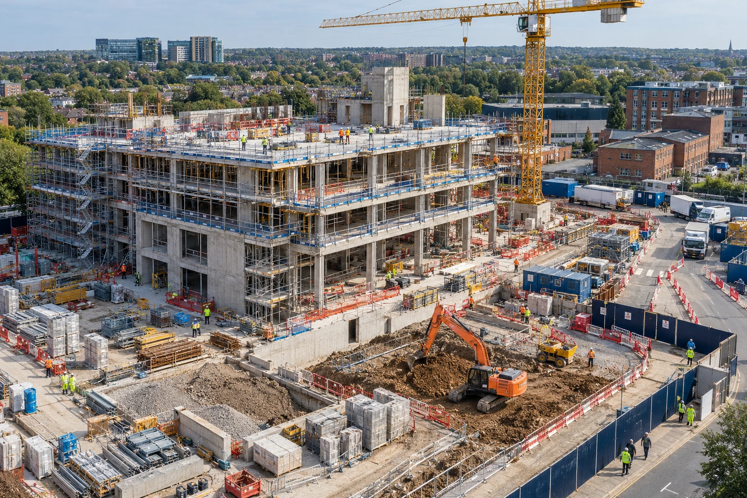

Specialist radiation work is not coordinated
All related inspection points 1 across 1 location
Construction site 1 inspection point
Practice in the SHAREhub environment. Browser practice is separate from workplace competence and formal learning records.
What are the key facts about radiation risk management?
Radiation risk management starts with source identification and ends with evidence that exposure is controlled.
Best-practice guide: Ionising and Non-Ionising Radiation Risk Management
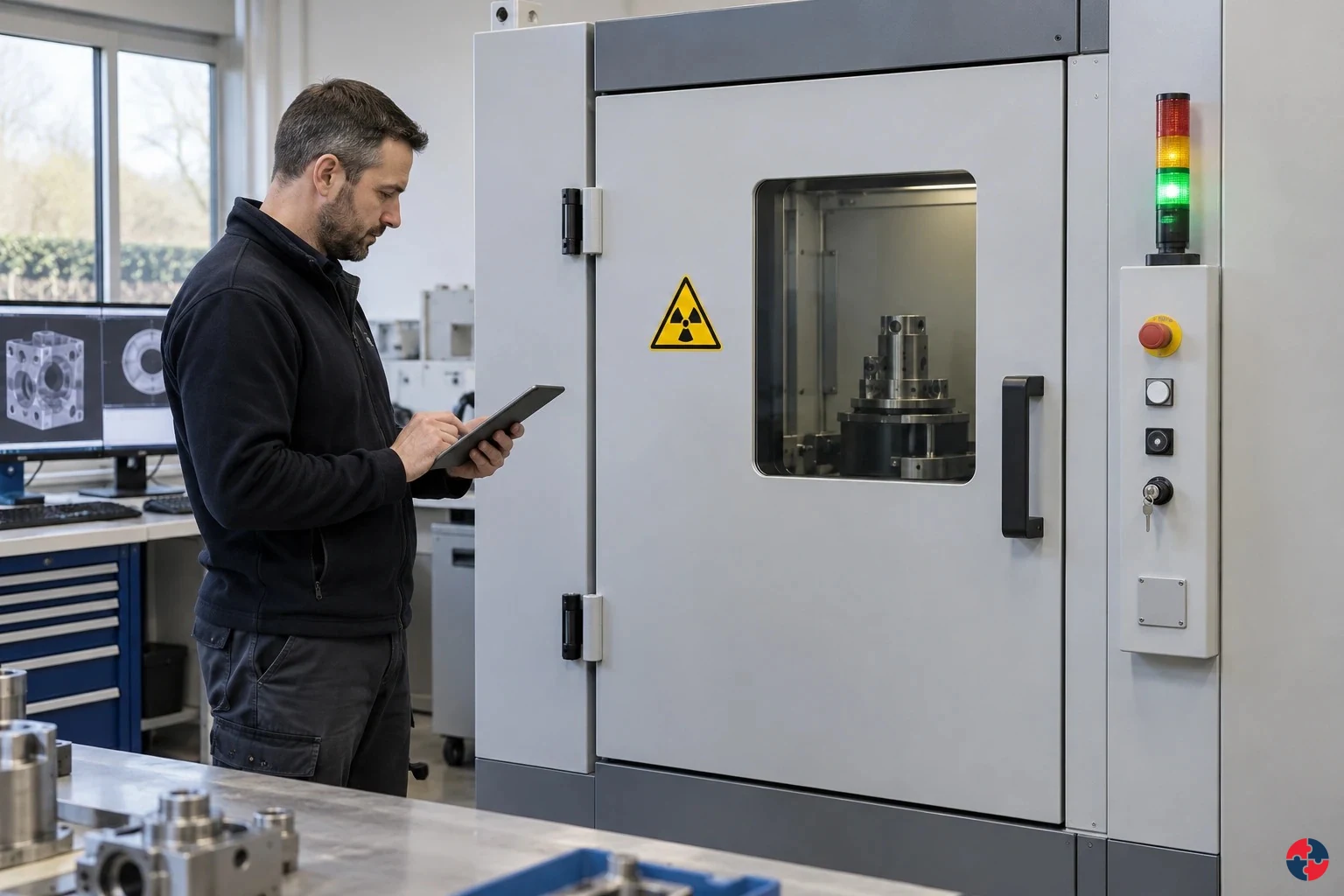
What are the key facts about radiation risk management?
Radiation risk management starts with source identification and ends with evidence that exposure is controlled. Radiation controls depend on the actual source and exposure route.
Turn ionising and non-ionising radiation risk management knowledge into a decision
Radiation controls depend on the actual source and exposure route. Ionising radiation, ultraviolet and laser radiation, electromagnetic fields and radon have different health mechanisms and legal controls. Keep contractor authorisation, permission route, assessment, local rules, permit, monitor results, alarm response and handback.
Contractor radiography near a live fuel terminal
The radiation protection supervisor controls the boundary and checks that no other crew enters while the source is active. The appointed competent radiographer/operator uses a suitable dose-rate monitor to confirm source retraction or de-energisation under the local arrangements; an alarm dosimeter alone is insufficient. With RPS oversight and RPA advice on the assessment and method, the site controller releases the area only after formal handback and the required evidence.
What health effects should be understood?
Radiation health effects depend on type, dose or intensity, body part, exposure duration and susceptibility.
Choose controls for ionising and non-ionising radiation risk management
Elimination: Avoid exposed source operation by using suitable external diagnostics and planned source-off servicing. Substitution: Evaluate a suitable non-ionising inspection method through quality and hazard review; another radiation type is not automatically harmless. Engineering controls: Securely isolate and prevent unintended source energisation for maintenance; restrict any authorised test area. Engineering controls: Restore specified shielding, enclosures, interlocks and warning functions and verify them using the approved test method. Administrative controls: Agree service/test scope, competent personnel, source control and release criteria with the radiation and operations owners. Read the full control table for qualifications, ownership and verification. Use the actual task and applicable jurisdiction; existing technical approval remains unchanged.
Use the full ionising and non-ionising radiation risk management lesson
Use the full topic for the decision points, practical examples, detailed control tables and source scope. HSE guidance applies to Great Britain; use HSENI for the Northern Ireland route and the source’s stated jurisdiction. A simplified guide does not change the page’s technical-review or approval status.

Read all six steps as text
1. What are the key facts about radiation risk management?
Radiation risk management starts with source identification and ends with evidence that exposure is controlled.
- Avoid / redesign: Avoid exposed source operation by using suitable external diagnostics and planned source-off servicing.
- Control the task: Securely isolate and prevent unintended source energisation for maintenance; restrict any authorised test area.
- Verify before release: If the hazardous step remains, keep it within the task assessment and select the stronger feasible remaining controls.
Radiation risk management starts with source identification and ends with evidence that exposure is controlled. Radiation controls depend on the actual source and exposure route.
Apply it: Which part of this plan would you check at the workface before authorising the task?
2. Turn ionising and non-ionising radiation risk management knowledge into a decision
Use the evidence to improve the actual work arrangement.
- Change at source: Avoid exposed source operation by using suitable external diagnostics and planned source-off servicing.
- Make it observable: If the hazardous step remains, keep it within the task assessment and select the stronger feasible remaining controls.
- Keep useful evidence: Keep contractor authorisation, permission route, assessment, local rules, permit, monitor results, alarm response and handback.
Radiation controls depend on the actual source and exposure route. Ionising radiation, ultraviolet and laser radiation, electromagnetic fields and radon have different health mechanisms and legal controls. Keep contractor authorisation, permission route, assessment, local rules, permit, monitor results, alarm response and handback.
Apply it: What evidence would show that the change improves this task, rather than only completing a record?
3. Contractor radiography near a live fuel terminal
Example: a practical application of the existing topic lesson.
- Setting: An industrial radiography contractor proposes night work near a fuel terminal access road while maintenance and security teams remain active.
- Act: Agree a specific permit, local rules, exclusion and warning arrangements; verify shielding, monitors and competent staffing before exposing the source.
- Hold / review: Withdraw, prevent entry and activate the approved contingency plan if the boundary fails, a monitor alarms or the source cannot be accounted for. Only specifically trained and authorised responders may approach, handle or attempt recovery.
The radiation protection supervisor controls the boundary and checks that no other crew enters while the source is active. The appointed competent radiographer/operator uses a suitable dose-rate monitor to confirm source retraction or de-energisation under the local arrangements; an alarm dosimeter alone is insufficient. With RPS oversight and RPA advice on the assessment and method, the site controller releases the area only after formal handback and the required evidence.
Apply it: At which point would you hold this example, and what evidence would allow it to proceed?
4. What health effects should be understood?
Connect the task, the route to the person and the control that can fail.
- Source / exposure: Radiation health effects depend on type, dose or intensity, body part, exposure duration and susceptibility.
- Possible consequence: High-dose ionising exposure can cause tissue injury; lower or prolonged dose increases later cancer risk.
- Control failure: Non-ionising effects must be assessed by source and route, not inferred from the word radiation alone.
Radiation health effects depend on type, dose or intensity, body part, exposure duration and susceptibility.
Apply it: Who could be exposed, including other trades, cleaners, maintainers and people nearby?
5. Choose controls for ionising and non-ionising radiation risk management
Start with the stronger practicable options and verify their effect.
- Elimination: Avoid exposed source operation by using suitable external diagnostics and planned source-off servicing.
- Substitution: Evaluate a suitable non-ionising inspection method through quality and hazard review; another radiation type is not automatically harmless.
- Engineering controls: Securely isolate and prevent unintended source energisation for maintenance; restrict any authorised test area.
- Engineering controls: Restore specified shielding, enclosures, interlocks and warning functions and verify them using the approved test method.
- Administrative controls: Agree service/test scope, competent personnel, source control and release criteria with the radiation and operations owners.
Elimination: Avoid exposed source operation by using suitable external diagnostics and planned source-off servicing. Substitution: Evaluate a suitable non-ionising inspection method through quality and hazard review; another radiation type is not automatically harmless. Engineering controls: Securely isolate and prevent unintended source energisation for maintenance; restrict any authorised test area. Engineering controls: Restore specified shielding, enclosures, interlocks and warning functions and verify them using the approved test method. Administrative controls: Agree service/test scope, competent personnel, source control and release criteria with the radiation and operations owners. Read the full control table for qualifications, ownership and verification. Use the actual task and applicable jurisdiction; existing technical approval remains unchanged.
Apply it: Which stronger option can be used here, and who will verify it before accepting the remaining risk?
6. Use the full ionising and non-ionising radiation risk management lesson
Follow the official sources below and retain the task-specific evidence.
- Official guidance: HSE - Introduction to radiation in the workplace
- Official guidance: HSE - Ionising radiation legal base
- Task decision: Keep contractor authorisation, permission route, assessment, local rules, permit, monitor results, alarm response and handback.
Use the full topic for the decision points, practical examples, detailed control tables and source scope. HSE guidance applies to Great Britain; use HSENI for the Northern Ireland route and the source’s stated jurisdiction. A simplified guide does not change the page’s technical-review or approval status.
Apply it: Which official source and full-topic section will you use to resolve the next decision?
Ionising radiation can damage tissue and raise later cancer risk; non-ionising sources do not all act alike. A sealed gauge, an X-ray unit, a welding arc, a laser, an MRI magnet and radon therefore need separate assessments. Time, distance and shielding help ionising control, while optical and EMF controls depend on source, wavelength or field, access and affected people. See UKHSA basic concepts of radiation; HSE - Non-ionising radiation.
Radiation is not one hazard
X-rays, radon, UV, lasers and EMFs behave differently and need different controls.
Ionising radiation needs specialist discipline
IRR17, RPAs, local rules, dose assessment and controlled areas may apply.
Non-ionising radiation can still harm
UV, lasers, infrared and EMFs can cause burns, eye injury, heating or interference effects.
Time, distance and shielding are core ideas
Exposure is reduced by limiting time near the source, increasing distance and using suitable shielding or enclosure.
Evidence is technical
Dose records, surveys, local rules, maintenance, signs, training and competent advice must be current and understood.
What does radiation risk management mean in plain English?
It means understanding the source, route of exposure, dose or intensity, who may be exposed and what prevents harm.
Radiation can be invisible and highly technical, so poor management often begins with not knowing the source is present. An XRF analyser, sealed gauge, industrial radiography source, radon-affected basement, UV curing lamp, welding arc, laser, induction heater or high-current electrical process may each need a different decision pathway.
The professional skill is to avoid both complacency and overreaction. Everyday low-risk office equipment usually needs little action, but high-power lasers, industrial radiography, radon in occupied basements or work with radioactive sources may need formal assessment, specialist advice, local rules, monitoring and emergency arrangements.
- What is the source? — Controls depend on the radiation type and equipment. X-ray set, radon, UV lamp, laser, EMF source.
- Who is exposed? — Workers, contractors, visitors and maintenance teams may differ. Contractor opens laser housing during repair.
- How strong and how long? — Dose or intensity depends on exposure conditions. Short industrial radiography exposure versus continuous radon.
- What controls exist? — Shielding, enclosure, distance and local rules need verification. Interlocked enclosure around X-ray cabinet.
- Who is competent? — Specialist interpretation is often needed. Radiation Protection Adviser or laser competent person.
How do the main radiation types differ?
The first learning step is to separate ionising radiation from non-ionising radiation and then understand the specific source.
Ionising radiation includes X-rays, gamma rays and particles such as alpha, beta and neutrons. It can ionise atoms and damage tissue or DNA, so dose control, shielding, source security, local rules and regulatory duties are central.
Non-ionising radiation includes ultraviolet, visible, infrared, radiofrequency, microwaves and electromagnetic fields. It does not have enough energy to ionise atoms, but high exposure can still harm skin, eyes or body tissues, or create specific EMF effects.
| Radiation type | Workplace examples | Primary control focus |
|---|---|---|
| X-rays and gamma rays | Industrial radiography, XRF analysers, gauges, inspection equipment. | Shielding, distance, access control, local rules and dose monitoring. |
| Alpha/beta/neutron radiation | Radioactive substances, nuclear work, sealed or unsealed sources. | Containment, source control, contamination control and monitoring. |
| Radon | Ground-floor rooms, basements, caves, tunnels, mines and underground work. | Testing, ventilation, remediation and RPA advice above action level. |
| Ultraviolet radiation | Welding arcs and UV curing are artificial sources; outdoor sunlight is a natural source assessed under general work-risk duties, not the artificial optical radiation regulations.HSE legal scope. | Enclosure, screens, PPE, skin/eye protection and work planning. |
| Lasers | Cutting, alignment, research, entertainment and medical/industrial equipment. | Classification, beam control, interlocks, eyewear and authorised users. |
| EMFs and radiofrequency | Induction heating, welding, broadcasting, MRI, power systems. | Assessment against action levels/ELVs and control of high-intensity sources. |
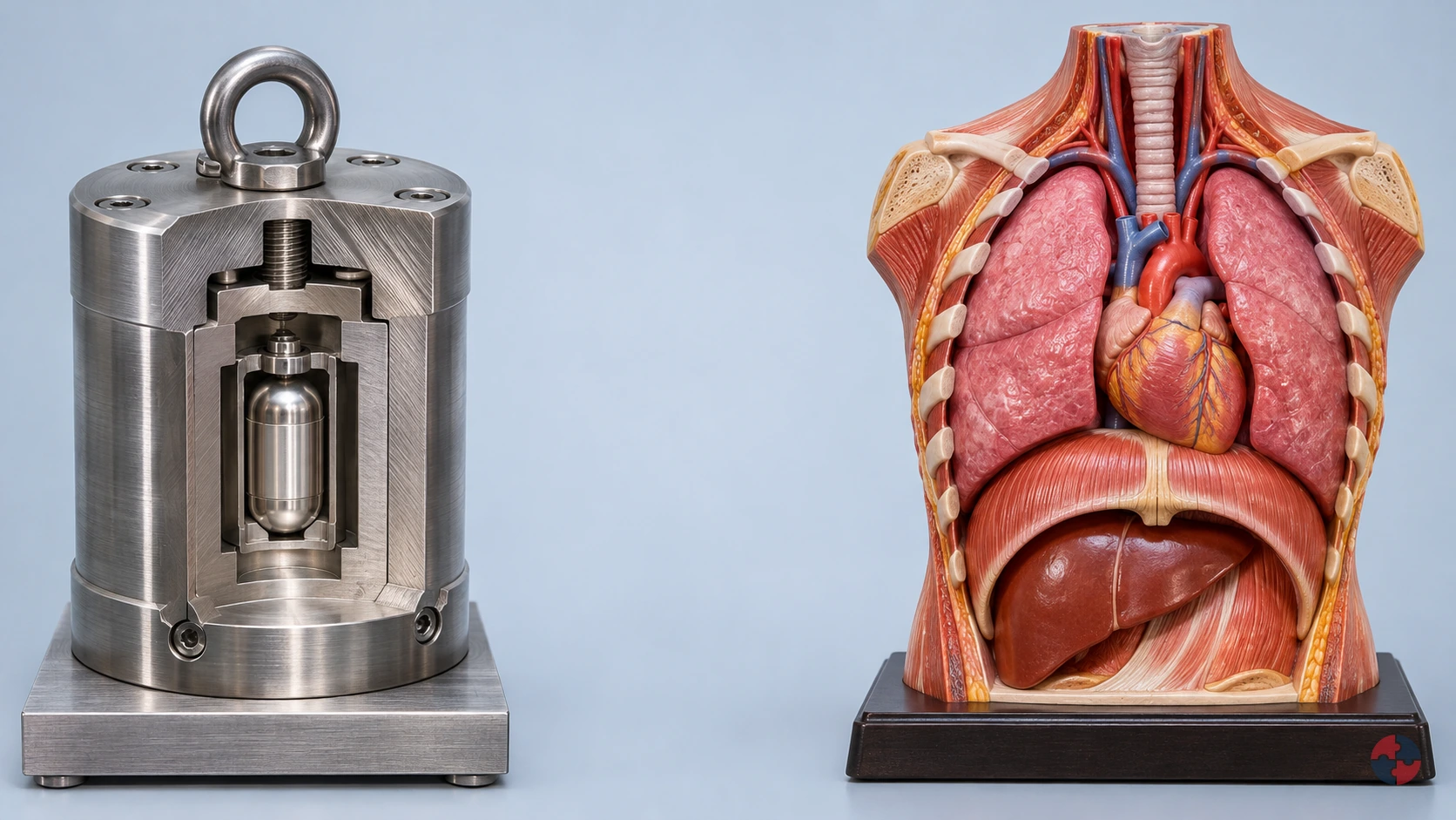
How should radiation type, distance, shielding and exposure route guide ionising-radiation controls?
Interactive radiation explainer · SHAREhub environment
Read all five radiation, shielding and body-effect explanations
Alpha
Typical sources: Alpha-emitting material Real example: radon and its radioactive decay products in buildings or underground workplaces. An unstable nucleus emits an alpha particle: two protons and two neutrons. The display holder is a teaching cutaway, not a radon container or an equipment design.
Travel and distance: A few centimetres in air Typical alpha particles rapidly lose energy in air. The illustrated track length is enlarged.
Penetration and exposure route: External: stops at the outer dead skin layer This applies to intact outer skin. Inside an airway or a wound, living cells have no such protective barrier.
Possible effects: A short, dense ionisation track can damage nearby cells. Inhaled radon decay products expose lung tissue and increase lung-cancer risk.
When radioactive material enters the body: Internal alpha: an airway deposit Illustrates an inhaled radioactive decay product near airway cells. Its short range concentrates energy locally; the exact organ dose depends on the radionuclide and where material is retained. 1 · A radioactive deposit is already inside The airway inset starts after an alpha-emitting radioactive decay product has been inhaled and deposited beside living cells. It can continue to emit radiation while it remains there. 2 · Follow a short track through local tissue The particle now starts inside the airway lining, rather than outside intact skin. It deposits energy densely over a short local track; short range does not make an internal alpha emitter harmless. 3 · Why an outside barrier cannot shield this deposit Paper outside the body cannot sit between the deposited emitter and adjacent cells. Containment and preventing intake address the exposure route; an exterior shield does not remove material already inside. 4 · Examine local cell interactions Nearby cells may receive energy that damages biological molecules, including DNA. Radon-related exposure can increase lung-cancer risk over time. The highlighted cells do not predict an immediate injury, a measured dose or a certain cancer outcome.
Shielding and control choices: Paper illustrates alpha stopping; preventing radioactive material entering the body is the crucial additional control. A paper barrier does not control airborne radon or contaminated dust.
Example sequence: 1 · See the source The teaching holder reveals alpha emissions. Radon-related exposure usually comes from radioactive gas and its decay products, not a visible glowing rock. 2 · Follow the short air track The particle deposits energy densely and stops over a short path. A few centimetres is a useful typical scale, not a clearance distance. 3 · Insert a barrier A sheet of paper stops the illustrated alpha track. The source remains radioactive even when no track reaches the body. 4 · Compare outside and inside Outside: the track stops at intact outer skin. Select internal exposure to see an airway deposit irradiate adjacent living cells. This body inset is a separate unshielded local comparison; it does not show the outcome of the selected barrier.
Compare barrier materials
- Air only: The short alpha track stops in air before distant tissue; close or internal exposure is a different situation. Material comparison only; thickness, energy and geometry are not calculated.
- Paper: Paper stops the illustrated alpha particles. Containment and control of intake still matter. Material comparison only; thickness, energy and geometry are not calculated.
- Plastic: Plastic stops the illustrated alpha particles. Containment and control of intake still matter. Material comparison only; thickness, energy and geometry are not calculated.
- Aluminium: Aluminium stops the illustrated alpha particles. Containment and control of intake still matter. Material comparison only; thickness, energy and geometry are not calculated.
- Lead: Lead stops the illustrated alpha particles. Containment and control of intake still matter. Material comparison only; thickness, energy and geometry are not calculated.
- Concrete: Concrete stops the illustrated alpha particles. Containment and control of intake still matter. Material comparison only; thickness, energy and geometry are not calculated.
- Water: Water stops the illustrated alpha particles. Containment and control of intake still matter. Material comparison only; thickness, energy and geometry are not calculated.
- Layered neutron barrier: Layered neutron barrier stops the illustrated alpha particles. Containment and control of intake still matter. Material comparison only; thickness, energy and geometry are not calculated.
Beta
Typical sources: Beta-emitting material Examples include radioactive tracers in laboratories and some industrial thickness gauges. This example shows beta-minus radiation: an electron emitted during nuclear decay. Generic teaching holder; real sources have purpose-designed containment and protection.
Travel and distance: Centimetres to metres, depending on energy Beta emitters have different energy distributions. Some beta particles travel several metres in air; low-energy examples travel much less.
Penetration and exposure route: Skin and superficial tissue; depth varies More energetic beta particles can reach living skin and the lens of the eye. Do not treat beta as harmless because it is less penetrating than gamma.
Possible effects: External beta exposure can injure skin or eyes at sufficient dose. Radioactive material inside the body can irradiate nearby tissue.
When radioactive material enters the body: Internal beta: a local deposit A simplified contaminated-tissue example. The same electron track now starts within the body. Actual distribution depends on the material; inhalation is not the only possible entry route. 1 · A beta emitter is already beside tissue This airway-cell example begins with radioactive material already deposited inside the body. Inhalation is one entry route; other materials can enter by swallowing or through wounds and distribute differently. 2 · Follow an electron from within the body The emitted beta electron scatters and loses energy in nearby tissue. Its finite local track depends on energy and tissue; the magnified drawing does not represent a fixed depth or organ dose. 3 · Separate outside shielding from internal exposure An external plastic panel cannot be placed between this deposited emitter and neighbouring cells. Preventing contamination entering the body matters; changing the outside panel cannot cancel an internal exposure. 4 · See local energy deposition and possible harm Interactions can affect cells close to the radioactive deposit. The amount and distribution of material, its emissions and retention determine exposure; a coloured track does not establish tissue injury or an individual cancer risk.
Shielding and control choices: Low atomic number plastic is a useful first comparison for beta shielding. Slowing beta particles can generate bremsstrahlung X-rays, especially in dense, high atomic number materials. A designed shield may need more than one layer.
Example sequence: 1 · See the beta emitter The electron starts at the nucleus. This is a beta-minus example; positron-emitting sources need additional explanation because annihilation photons are also produced. 2 · Follow the changing track Interactions scatter and slow the electron. Its travel depends strongly on starting energy, so there is no single range shared by all beta sources. 3 · Compare plastic and lead Plastic absorbs the illustrated beta track with less bremsstrahlung than a comparable high atomic number material. Lead is not an automatic first choice. 4 · See superficial energy deposition The enlarged skin section shows a finite electron track in tissue. Internal contamination places the emitter beside living cells; it changes the exposure route. This body inset is a separate unshielded local comparison; it does not show the outcome of the selected barrier.
Compare barrier materials
- Air only: Longer beta tracks may reach exposed skin; range changes with energy. Material comparison only; thickness, energy and geometry are not calculated.
- Paper is not a universal beta barrier: The example passes through paper. Very low energy beta emitters behave differently. Material comparison only; thickness, energy and geometry are not calculated.
- Low atomic number first layer: Plastic illustrates electron stopping while limiting bremsstrahlung production. Material comparison only; thickness, energy and geometry are not calculated.
- Electron stopping with a secondary-radiation check: Aluminium can stop beta particles; energy and shield thickness determine the result. Bremsstrahlung still needs consideration. Material comparison only; thickness, energy and geometry are not calculated.
- Stopping electrons can create X-rays: High atomic number lead can increase bremsstrahlung production. A real beta shield may start with plastic and use another layer for photons. Material comparison only; thickness, energy and geometry are not calculated.
- Concrete: Beta tracks lose energy in material; a substantial barrier can absorb the electrons. Secondary photons and real construction still need assessment. Material comparison only; thickness, energy and geometry are not calculated.
- Water: Beta tracks lose energy in material; a substantial barrier can absorb the electrons. Secondary photons and real construction still need assessment. Material comparison only; thickness, energy and geometry are not calculated.
- Layered barrier: Beta tracks lose energy in material; a substantial barrier can absorb the electrons. Secondary photons and real construction still need assessment. Material comparison only; thickness, energy and geometry are not calculated.
Gamma
Typical sources: Gamma-emitting radioactive source Industrial radiography uses shielded radioactive sources to inspect components such as pipe welds. A nucleus releases a photon as it moves to a lower energy state. Generic capsule in an illustrative cutaway; real radiography sources remain within engineered systems.
Travel and distance: Long-range: attenuates rather than ending at one distance Photons can cross substantial air gaps. Distance changes exposure but does not create a universal safe boundary.
Penetration and exposure route: Can reach deep organs or pass through the body Some photons interact and deposit energy; others pass through. Penetration depends on energy, tissue and geometry.
Possible effects: Energy deposited in tissue can damage cells and DNA. Risk depends on dose and exposed tissue; sufficiently high doses can also cause tissue injury.
Shielding and control choices: Lead and appropriately designed concrete barriers reduce photon transmission. Shielding attenuates the beam. It is misleading to show one standard sheet as stopping every gamma photon.
Example sequence: 1 · See a continuing radioactive emission The capsule emits gamma photons as radioactive decay proceeds. Turning off an electrical supply does not stop radioactive decay. 2 · Follow photons across the scene Unlike the short alpha path, photon transmission falls progressively. The scene is compressed and provides no safe-distance calculation. 3 · Reduce the transmitted beam Absorption and scattering in the barrier leave fewer illustrated forward tracks. Some transmission remains: real design checks also consider scattered radiation and openings. 4 · See interactions at different depths A penetrating photon can transfer energy inside the body. The highlighted points represent interactions, not a prediction of injury or cancer. This body inset is a separate unshielded local comparison; it does not show the outcome of the selected barrier.
Compare barrier materials
- Unshielded path: Photons can continue across the scene and into tissue. Distance and time influence exposure. Material comparison only; thickness, energy and geometry are not calculated.
- Paper gives little protection in this example: The illustrated photon beam largely continues. Paper is not an appropriate photon protection strategy. Material comparison only; thickness, energy and geometry are not calculated.
- Plastic: Some attenuation occurs, but this illustrative layer leaves substantial photon transmission. Energy and actual thickness matter. Material comparison only; thickness, energy and geometry are not calculated.
- Aluminium: Some attenuation occurs, but this illustrative layer leaves substantial photon transmission. Energy and actual thickness matter. Material comparison only; thickness, energy and geometry are not calculated.
- Dense photon barrier: Lead reduces forward transmission through absorption and scattering; some photons still transmit or scatter. Material comparison only; thickness, energy and geometry are not calculated.
- Structural photon shielding: A designed concrete barrier can reduce the field. Composition, thickness, joints and openings affect its performance. Material comparison only; thickness, energy and geometry are not calculated.
- Water also attenuates photons: A sufficient path through water reduces transmission. The small illustrated layer does not establish a protective thickness. Material comparison only; thickness, energy and geometry are not calculated.
- Assess the photon component separately: A neutron-oriented composite is not automatically a complete photon shield. Assess the materials and photon energies actually present. Material comparison only; thickness, energy and geometry are not calculated.
X-rays
Typical sources: Energised X-ray tube Medical and dental imaging, security screening, cabinet inspection and handheld X-ray fluorescence analysis. Electrons accelerated within a tube strike a target and produce X-ray photons. The beam appears only during the illustrated energised exposure. This is not a radioactive source capsule.
Travel and distance: No single stopping distance in air X-rays are photons. Beam energy, filtration, direction and scatter all affect transmission.
Penetration and exposure route: Can penetrate the body; absorption differs between tissues Differences in transmission allow diagnostic images. The examination uses a controlled, justified exposure.
Possible effects: X-rays can damage cells, so exposures are controlled. A normal diagnostic X-ray does not leave radiation stored inside a person or make them radioactive.
Shielding and control choices: Purpose-designed shielding such as lead or concrete reduces the primary and scattered X-ray field. X-rays are not always less penetrating than gamma rays: photon energy, not the name alone, governs their interactions.
Example sequence: 1 · Energise the teaching tube An electron beam meets the target; X-ray photons leave the tube window. A normal diagnostic tube produces its beam while energised. 2 · Follow a directed beam and scatter The illustrated beam travels through air. Some interactions redirect photons; this is why the forward beam alone does not describe every exposure. 3 · Place a designed photon barrier The barrier reduces transmission. Real installations account for equipment output, workload, occupants and scatter, with suitable expert advice. 4 · Compare tissue transmission Some photons cross the body and some interact. The beam ends when the diagnostic exposure ends; the patient does not retain the X-ray beam. This body inset is a separate unshielded local comparison; it does not show the outcome of the selected barrier.
Compare barrier materials
- Unshielded path: Photons can continue across the scene and into tissue. Distance and time influence exposure. Material comparison only; thickness, energy and geometry are not calculated.
- Paper gives little protection in this example: The illustrated photon beam largely continues. Paper is not an appropriate photon protection strategy. Material comparison only; thickness, energy and geometry are not calculated.
- Plastic: Some attenuation occurs, but this illustrative layer leaves substantial photon transmission. Energy and actual thickness matter. Material comparison only; thickness, energy and geometry are not calculated.
- Aluminium: Some attenuation occurs, but this illustrative layer leaves substantial photon transmission. Energy and actual thickness matter. Material comparison only; thickness, energy and geometry are not calculated.
- Dense photon barrier: Lead reduces forward transmission through absorption and scattering; some photons still transmit or scatter. Material comparison only; thickness, energy and geometry are not calculated.
- Structural photon shielding: A designed concrete barrier can reduce the field. Composition, thickness, joints and openings affect its performance. Material comparison only; thickness, energy and geometry are not calculated.
- Water also attenuates photons: A sufficient path through water reduces transmission. The small illustrated layer does not establish a protective thickness. Material comparison only; thickness, energy and geometry are not calculated.
- Assess the photon component separately: A neutron-oriented composite is not automatically a complete photon shield. Assess the materials and photon energies actually present. Material comparison only; thickness, energy and geometry are not calculated.
Neutrons
Typical sources: Neutron-emitting source Examples include specialist research, nuclear facilities and neutron gauges; an americium–beryllium capsule is one neutron-source type. Some high-energy medical accelerators produce secondary neutrons. Nuclear reactions can release uncharged neutrons. This teaching example represents a contained neutron source. Generic source representation; mixed neutron and gamma fields require specialist assessment.
Travel and distance: Energy-dependent; potentially highly penetrating Neutrons scatter and change energy. Neither a fixed air range nor a single lead sheet describes their behaviour.
Penetration and exposure route: Can irradiate deep tissue Neutrons transfer energy through nuclear interactions, producing charged particles that deposit energy locally.
Possible effects: Neutron interactions can damage tissue and increase cancer risk. Biological effect varies with neutron energy; counting tracks alone does not compare doses between radiation types.
Shielding and control choices: Hydrogen-rich material slows fast neutrons; a capture component can remove slowed neutrons. A designed barrier also considers photons. Capture and other interactions can produce secondary gamma radiation. Moderating a neutron is not the same as removing all radiation.
Example sequence: 1 · See an uncharged neutron leave the source The neutron starts from a nuclear process, not from the electron beam of an ordinary diagnostic X-ray tube. Real sources may emit gamma radiation as well. 2 · Follow scattering and changing energy A neutron can travel well beyond the short alpha path. Collisions can deflect it and reduce its energy, rather than simply making it vanish. 3 · Slow, capture and consider secondary photons Hydrogen-rich shielding illustrates moderation, followed by a capture component. A secondary gamma track reminds learners that the whole field must be assessed. 4 · See energy transferred to tissue nuclei An interaction creates an energetic charged recoil or reaction product. The short recoil track represents local energy deposition, not a visible wound or a dose estimate. This body inset is a separate unshielded local comparison; it does not show the outcome of the selected barrier.
Compare barrier materials
- No moderating or capture barrier: Neutrons can reach tissue. Scattering and energy changes make a universal air-range claim unsuitable. Material comparison only; thickness, energy and geometry are not calculated.
- Paper: This is not a complete fast-neutron protection strategy. A material effective for photons may perform poorly for neutrons. Material comparison only; thickness, energy and geometry are not calculated.
- Aluminium: This is not a complete fast-neutron protection strategy. A material effective for photons may perform poorly for neutrons. Material comparison only; thickness, energy and geometry are not calculated.
- Lead: This is not a complete fast-neutron protection strategy. A material effective for photons may perform poorly for neutrons. Material comparison only; thickness, energy and geometry are not calculated.
- Hydrogen-rich plastic moderates: Collisions with hydrogen help slow fast neutrons. Moderation alone does not guarantee capture or control secondary radiation. Material comparison only; thickness, energy and geometry are not calculated.
- Concrete can shield mixed fields: Hydrogen content and composition matter for neutrons; the design must also account for secondary photons. Material comparison only; thickness, energy and geometry are not calculated.
- Water illustrates moderation: Hydrogen helps slow neutrons. Some interactions produce secondary gamma radiation; a real barrier is assessed as a whole. Material comparison only; thickness, energy and geometry are not calculated.
- Moderation plus capture: Hydrogen-rich material slows fast neutrons and a capture component removes some slowed neutrons. Secondary gamma shielding may also be needed. Material comparison only; thickness, energy and geometry are not calculated.
Sources and how to interpret the model
Original teaching geometry and animation. The reference diagram is a layout inspiration; no third-party illustration has been copied. Coloured tracks, counts, timing and materials illustrate mechanisms rather than exposure measurements. This activity is not a radiation protection assessment or a design specification.
- Health and Safety Executive: Ionising radiation: protecting workers and others
- UK Health Security Agency: Basic concepts of radiation
- UK Health Security Agency: Living with radiation
- Health and Safety Executive: Radon in the workplace
- Health and Safety Executive: Introduction to radiation in the workplace
- Health and Safety Executive: Work with ionising radiation that requires registration
- Health and Safety Executive: Industrial radiography – managing radiation risks (IRP1)
- NHS: X-ray
- North Tees and Hartlepool NHS Foundation Trust: My X-ray Examination – What You Need to Know about Radiation and the Risks Involved
- Office for Nuclear Regulation: Shielding and dose rate safety assessment of transport packages (NS-TAST-GD-100)
- Office for Nuclear Regulation: Criticality safety assessment of transport packages (NS-TAST-GD-097)
- Health and Safety Executive: Radiation Protection Advisers
- Health and Safety Executive: Dose monitoring, assessment and recording
- UK Health Security Agency: Accidental neutron exposure in a medical setting: a case study
- Office for Nuclear Regulation: Step 4 assessment of radiological protection for the UK EPR (ONR-GDA-AR-11-025)
What learner comparisons matter for radiation risk management?
Diploma-level understanding depends on comparing concepts that are often confused, then linking them to real controls.
Radiation questions become much easier when the learner can hold the main contrasts in their head. Ionising and non-ionising radiation are different hazards; deterministic and stochastic effects are different health-effect models; controlled and supervised areas are different levels of management control; and an RPA and RPS have different roles in the system.
The practical test is whether the learner can take the concept and apply it to a workplace decision. For example, radon in a basement is managed through measurement and remediation; an industrial radiography job needs exclusion and specialist control; a UV welding arc needs screens and eye protection; a high-power laser needs beam control and authorised users.
- Ionising radiation — Radiation with enough energy to ionise atoms and damage DNA or tissue. Industrial radiography, X-ray equipment, sealed gauges and radon need specialist assessment, source control and dose management.
- Non-ionising radiation — Radiation that does not ionise atoms but can still cause heating, burns, eye injury or other effects. UV welding, lasers, infrared sources and EMF equipment need screens, enclosures, exclusion, eyewear or exposure assessment.
- Deterministic effects — Tissue effects that normally have a threshold; severity increases with dose above that threshold. Skin injury, cataracts or acute radiation effects make high-dose prevention and investigation essential.
- Stochastic effects — Effects where the probability increases with dose, without a simple threshold. Long-term cancer-risk prevention supports ALARP exposure, dose records and conservative control decisions.
- Dose, time, distance and shielding — Dose is affected by exposure duration, proximity and barriers between the source and person. A radiography job reduces dose by minimising time near source, increasing distance and using suitable shielding/exclusion.
- Controlled and supervised areas — Designated areas used to control access and exposure where ionising radiation work requires it. A controlled area may need local rules, signage, authorised access and RPS oversight.
- RPA and RPS roles — The RPA gives competent radiation protection advice; the RPS supervises local rules where appointed. A manager should not invent local rules without RPA advice or leave RPS duties unclear.
- Radon — Naturally occurring radioactive gas that can build up in occupied workplaces. Basements, cellars, tunnels and radon-affected areas may need measurement, remediation and review.
- UV, lasers and EMFs — Non-ionising sources with different exposure mechanisms. A welding bay, Class 4 laser or high-current process needs source-specific assessment, barriers and training.
What health effects should be understood?
Radiation health effects depend on type, dose or intensity, body part, exposure duration and susceptibility.
High-dose ionising exposure can cause tissue injury; lower or prolonged dose increases later cancer risk. Non-ionising effects must be assessed by source and route, not inferred from the word radiation alone. UKHSA basic concepts of radiation; HSE - Non-ionising radiation.
For ionising radiation, the concern can include acute tissue damage at high doses and long-term stochastic effects such as cancer risk. For non-ionising radiation, the concern may be skin burns, eye injury, cataracts, arc eye, thermal effects, photosensitivity or laser retinal injury.
A good risk assessment does not list every possible disease. It connects the realistic exposure mechanism to the task: the eye close to a laser beam, skin exposed to UV, the body near high EMF fields, or workers occupying a radon-affected basement.
- Ionising radiation dose — Cell damage, cataracts, skin effects or increased cancer risk. Dose restriction, RPA advice, monitoring and medical surveillance where required.
- Radon inhalation — Increased lung cancer risk from long-term exposure. Measure, remediate and monitor occupied areas.
- UV exposure — Sunburn, arc eye, skin damage and increased skin cancer risk. Screens, clothing, eye protection and work planning.
- Infrared — Skin burns and cataract risk near intense sources. Distance, shielding and eye/skin protection.
- Laser beam — Severe eye injury or skin burn. Beam enclosure, interlocks and laser safety controls.
- High EMF exposure — Sensory or health effects depending on frequency and intensity. Assessment, exclusion zones and equipment controls.
How should radiation risks be assessed?
Radiation assessment should be source-specific and should identify the realistic exposure scenario.
The assessor should identify equipment, source type, energy/intensity, operating mode, shielding, beam path, access points, maintenance tasks, abnormal situations, contractors, vulnerable groups, warning systems and failure modes. Radiation risk often changes during maintenance, alignment, source change, breakdown or unusual contractor work.
For ionising radiation, specialist advice is normally central. For non-ionising radiation, many workplaces can use manufacturer information and HSE guidance, but high-power lasers, intense optical sources and specialist EMF sources still need competent assessment.
- Is the source ionising or non-ionising? — Determines legal framework and specialist advice. Source inventory and equipment specification.
- Is exposure routine or accidental? — Controls must cover normal and foreseeable abnormal work. Task analysis and maintenance review.
- Can exposure be engineered out? — Design control is stronger than behaviour control. Shielding, enclosure and interlock evidence.
- Who needs access? — Unauthorised access may create serious risk. Local rules, barriers and access list.
- Is monitoring needed? — Dose or intensity may need measurement. Dose records, survey report or radon test.
- What happens if controls fail? — Emergency arrangements may be required. Emergency plan and drill/debrief.
How is ionising radiation controlled?
Ionising radiation control normally combines justification, restriction, dose limits, competent advice, local rules and monitoring.
The classic practical controls are time, distance and shielding. Reduce the time people spend near the source, increase distance from the source, and use shielding or containment suited to the radiation type. But mature control goes further: source security, interlocks, local rules, controlled/supervised areas, dosimetry, appointed roles, maintenance and emergency arrangements may all be needed. See L121 - Working with ionising radiation; Ionising Radiations Regulations 2017.
For Diploma-level understanding, remember that PPE is not the first answer. The better answer usually begins with design, source containment, remote handling, shielding, engineered interlocks, restricted access and competent radiation protection advice.
| Control | How it works | Evidence |
|---|---|---|
| Time | Minimises duration of exposure. | Planned task time and dose planning. |
| Distance | Uses separation to reduce exposure. | Exclusion zone and work layout. |
| Shielding | Places suitable material between source and person. | Shielding specification and survey. |
| Containment/source security | Prevents spread, loss or unauthorised access. | Source register and security checks. |
| Local rules | Set required working arrangements for controlled areas. | Local rules and worker acknowledgement. |
| Dose monitoring | Checks actual exposure where required. | Dosimetry and dose records. |
| Medical surveillance | Supports classified persons where required. | Appointed doctor and health record arrangements. |
How is non-ionising radiation controlled?
Non-ionising controls depend on the source: optical, laser, UV, infrared, radiofrequency, microwave or EMF.
Non-ionising radiation can be wrongly dismissed because it does not ionise atoms. That is a mistake. A welding arc can injure eyes and skin; a high-power laser can permanently damage vision; infrared from hot processes can injure eyes; high EMF fields can require specific assessment and exclusion zones. See Control of Artificial Optical Radiation at Work Regulations 2010; Control of Artificial Optical Radiation at Work Regulations (Northern Ireland) 2010; Control of Electromagnetic Fields at Work Regulations 2016; Control of Electromagnetic Fields at Work Regulations (Northern Ireland) 2016.
Controls should follow the same prevention logic: choose safer equipment, enclose or shield the source, interlock access, separate people, reduce exposure time, control maintenance access, train workers and use suitable PPE where needed.
| Source | Control focus | Example evidence |
|---|---|---|
| UV welding arc | Screens, eye protection, skin cover and exclusion of bystanders. | Welding bay inspection and PPE checks. |
| Laser | Classification, beam path, interlocks and authorised users. | Laser risk assessment and maintenance controls. |
| Infrared/hot process | Distance, shielding and eye/skin protection. | Furnace/glass process controls. |
| EMF | Assessment against action levels and exposure limit values. | EMF assessment and equipment data. |
| Microwave/radiofrequency | Enclosure, guarding and leakage checks. | Maintenance and inspection records. |
How should radon be managed?
Radon is a naturally occurring radioactive gas that can become a workplace radiation risk in buildings and underground areas.
Radon is invisible and cannot be detected by smell. For relevant occupied workplaces, measure the annual average concentration. An annual average above 300 becquerels per cubic metre (Bq/m³) triggers the GB IRR17 route and the separate NI notification route; it is a decision trigger, not permission to ignore risk below it. Reduce exposure, obtain competent advice where required, and remeasure after remediation. HSE workplace radon; HSENI NI permission overview.
Above that annual-average trigger, the employer should restrict exposure, use competent radiation advice and decide the GB or NI notification route. Ventilation, a sub-floor sump or changed occupancy needs design, commissioning and remeasurement; a lower reading does not excuse failure to assess relevant occupied space.
- Screen location — Use radon maps or definitive dataset where needed. Radon risk review.
- Measure — Use suitable passive detectors in relevant occupied areas. Radon measurement report.
- Interpret — Compare annual average to action level and risk profile. Competent review and decision record.
- Control — Improve ventilation, install sump or change occupancy where needed. Remediation record.
- Review — Remeasure after changes and periodically. Review schedule and results.
Make the measurement decision traceable
Occupied below-ground workplaces may need measurement regardless of the above-ground radon map. Identify occupied locations → obtain representative measurements → interpret the annual average with competent advice → check the applicable notification/remediation route → implement controls → check effectiveness promptly and obtain the required subsequent annual-average assessment. A short reading is not automatically an annual average. Keep the accurate 300 Bq/m³ workplace annual-average trigger and the distinct NI route.
Who needs competent radiation advice?
Competent advice is essential where the risk is specialist, legally controlled or technically uncertain.
For ionising radiation, a Radiation Protection Adviser is a defined role under IRR17. Organisations may also need Radiation Protection Supervisors, appointed doctors, dosimetry services, laser safety advice, occupational hygienists or equipment specialists depending on the hazard. See L121 - Working with ionising radiation.
The presence of a specialist does not remove line management responsibility. Managers still need to make sure advice is understood, actions are funded, controls are implemented and workers follow local rules.
- Radiation Protection Adviser — Ionising radiation work and IRR17 compliance advice. Risk assessment advice, local rules and dose-control guidance.
- Radiation Protection Supervisor — Supervises local rules in controlled/supervised areas. Local implementation and worker checks.
- Appointed doctor — Medical surveillance for classified persons. Medical surveillance records and fitness advice.
- Laser competent person — High-power laser risk assessment and controls. Laser controls and authorised-user rules.
- Occupational hygienist/physicist — Measurement and technical interpretation. Survey report and recommendations.
What monitoring and records may be needed?
Monitoring should prove exposure is controlled and identify deterioration before harm occurs. See HSE - Ionising radiation legal base.
- Personal dosimetry — Assesses individual ionising radiation exposure. Dose records and review.
- Area survey — Checks dose rates or intensity around equipment. Survey map and action list.
- Radon measurement — Assesses annual average radon levels. Detector report and remediation review.
- EMF assessment — Checks exposure against action levels/ELVs where relevant. Assessment report and exclusion zones.
- Optical/laser review — Checks beam control, eyewear, interlocks and access. Inspection and maintenance records.
- Health/medical surveillance — Supports classified persons or health concerns. Health records with confidentiality controls.
When are emergency arrangements needed?
Emergency planning depends on credible release, loss of shielding, overexposure, contamination, source loss or public impact.
A damaged gauge may need local source-control instructions; higher-hazard work can trigger distinct GB or NI radiation emergency regulations when the legal thresholds apply. Do not assume the GB route covers NI. GB REPPIR 2019; NI REPPIR 2019
Emergency instructions should be short, role-based and practised enough that workers do not improvise around invisible hazards.
- Industrial radiography source fails to retract — Evacuate/exclude, contact competent radiation lead. Dose assessment and investigation.
- Gauge damaged by vehicle — Isolate area and prevent access. RPA advice and regulator/insurer notification where needed.
- Laser eye exposure suspected — Stop use and arrange medical assessment route. Investigate controls and authorisation.
- Radon remediation fan fails — Review occupancy and monitor levels. Repair and remeasure.
- Contamination suspected — Control spread and seek specialist advice. Decontamination and exposure review.
Local contingency planning and reporting
Foreseeable radiography failures require rehearsed local contingency arrangements even where REPPIR does not apply. Keep people clear; no untrained approach or recovery. The responsible GB dutyholder checks IRR notification and the separate RIDDOR dangerous-occurrence categories for specified source/generator failures. Use HSENI’s separate NI reporting route for NI work; REPPIR applicability does not replace these checks.
HSE: industrial radiography roles and monitoring · HSE: ionising-radiation protection and reporting
What evidence shows radiation risk is managed?
Evidence should show source control, competent advice, monitoring, training and review.
Evidence should be read as a chain rather than a filing exercise. A single record rarely proves good management on its own; stronger assurance comes from matching records, observations, interviews, trends and verified action closure.
- Source inventory — Shows what sources exist and where. Is it current after procurement/disposal?
- Risk assessment — Links source to people, exposure and controls. Does it cover maintenance and abnormal work?
- RPA advice/local rules — Shows specialist control requirements. Are actions implemented?
- Dose/radon/survey records — Shows exposure is measured where needed. Are results reviewed and acted on?
- Training records — Shows workers understand local rules and warning signs. Is training role-specific?
- Maintenance/interlock checks — Shows engineered controls remain reliable. Are defects escalated?
- Emergency drills — Shows response arrangements work. Are debrief actions closed?
How does radiation risk appear in real workplaces?
The source determines the decision: an X-ray analyser, radon-affected room, exposed laser beam, welding arc and electromagnetic field need different assessments and controls. In each example, first identify the source and people exposed, then check the correct specialist and legal route before deciding that work may proceed.
A boundary around a radiography source is a different control from screening welding ultraviolet light or assessing an electromagnetic field near an implanted device. A site controller should therefore ask what source will be used, who may enter the area, what monitor or enclosure proves control, and who can stop or release the task. HSE ionising-radiation guidance applies to the first example; the optical and EMF cases need their own source-specific route.
- Scrap yard uses handheld XRF analyser — Ionising beam and scatter during metal analysis; check whether the analyser uses an X-ray tube or a radionuclide source. Train users, prevent pointing at body, maintain device and control public access.
- Basement office in radon-affected area — Long-term radon exposure. Measure, consult advice if high and remediate/ventilate.
- Warehouse repairs laser scanner housing — Embedded laser exposure during maintenance. Authorise maintenance and control access to open beam path.
- Fabrication bay with welding arcs — UV exposure to nearby workers. Use screens, PPE, bystander control and supervision.
- Healthcare contractor works near MRI/EMF sources — EMF effects and implanted-device concerns. Pre-job assessment, exclusions and specialist advice.
Contractor radiography near a live fuel terminal
Task, material and setting: An industrial radiography contractor proposes night work near a fuel terminal access road while maintenance and security teams remain active.
People and prerequisites: The site controller and contractor radiation protection adviser check source, exposure boundary, work permission, neighbouring occupancy and emergency contacts.
Before: Agree a specific permit, local rules, exclusion and warning arrangements; verify shielding, monitors and competent staffing before exposing the source.
During: The radiation protection supervisor controls the boundary and checks that no other crew enters while the source is active.
Hold or stop: Withdraw, prevent entry and activate the approved contingency plan if the boundary fails, a monitor alarms or the source cannot be accounted for. Only specifically trained and authorised responders may approach, handle or attempt recovery.
Completion and hand back: The appointed competent radiographer/operator uses a suitable dose-rate monitor to confirm source retraction or de-energisation under the local arrangements; an alarm dosimeter alone is insufficient. With RPS oversight and RPA advice on the assessment and method, the site controller releases the area only after formal handback and the required evidence.
Evidence: Keep contractor authorisation, permission route, assessment, local rules, permit, monitor results, alarm response and handback.
MRI access has a persistent-field branch
MRI assessment must address ferromagnetic projectile attraction as well as implanted-device susceptibility. Screen people, tools and equipment before controlled entry under specialist arrangements. For superconducting and permanent-magnet MRI systems the static field may remain when scanning stops. Induction equipment needs its own source/task assessment; the controls are not interchangeable and ordinary PPE is not a generic field shield.
Which legal duties apply to radiation risk management?
Choose the legal route only after identifying the source and work location. HSE’s ionising-radiation legal base explains the Great Britain IRR17 route; Northern Ireland has its own ionising-radiation regime. Artificial optical radiation and electromagnetic fields have separate rules; a laser, welding arc or EMF source should not be classified as an ionising source merely because all are called radiation.
Before a contractor brings a source on site, the responsible manager and competent radiation adviser should resolve the permission route, neighbouring work, exclusion boundary, local rules and credible emergency arrangements. If the source type, location or controlled boundary changes, hold the task and revisit the relevant GB or NI instrument rather than reusing the previous permit. The table below separates these routes.
| Jurisdiction | Duty route | Practical decision and evidence |
|---|---|---|
| Great Britain | Ionising Radiations Regulations 2017 | Classify the work and permission route; use a radiation protection adviser, restriction of exposure, local rules and dose arrangements where required. Sources: Ionising Radiations Regulations 2017; HSE - Ionising radiation legal base; HSE notification, registration and consent |
| Northern Ireland | Ionising Radiations Regulations (Northern Ireland) 2017 (SR 2017/229) | Follow HSENI’s separate notification, registration and consent route and obtain NI-specific competent advice before work. Official text and HSENI permission guidance: NI Ionising Radiations Regulations 2017; HSENI ionising radiation regulations |
| Great Britain | Artificial optical radiation and electromagnetic-field regulations | Assess relevant UV/laser and EMF sources under their separate GB regimes; check exposure limits, vulnerable workers and source-specific controls. Sources: Control of Artificial Optical Radiation at Work Regulations 2010; Control of Electromagnetic Fields at Work Regulations 2016 |
| Northern Ireland | Artificial optical radiation regulations (NI) 2010 | Apply the NI artificial-source route to UV, laser and intense optical work; natural sunlight is excluded from this specific regime. Sources: Control of Artificial Optical Radiation at Work Regulations (Northern Ireland) 2010 |
| Northern Ireland | Electromagnetic fields regulations (NI) 2016 | Assess relevant EMF work and affected people under the separate NI instrument; do not substitute GB law. Sources: Control of Electromagnetic Fields at Work Regulations (Northern Ireland) 2016 |
| Great Britain | Radiation emergency regulations 2019, where thresholds apply | Use the GB REPPIR route for qualifying radiation emergencies and L126 guidance. Sources: Radiation (Emergency Preparedness and Public Information) Regulations 2019; L126 - REPPIR 2019 ACOP and guidance |
| Northern Ireland | Radiation emergency regulations (NI) 2019, where thresholds apply | Use the NI REPPIR route; HSENI has approved L126 for NI use. Sources: Radiation (Emergency Preparedness and Public Information) Regulations (Northern Ireland) 2019; HSENI approval of L126 for NI use |
| Great Britain | Health and Safety at Work etc. Act 1974 | Apply GB general risk duties alongside the radiation-specific regime and competent advice. Official text: Health and Safety at Work etc. Act 1974 |
| Northern Ireland | Health and Safety at Work (Northern Ireland) Order 1978 | Apply NI general risk duties alongside the separate NI radiation regime and HSENI permission route. Official text: Health and Safety at Work (Northern Ireland) Order 1978 |
How could Crownway Fuels Ltd manage this in practice?

Crownway Fuels Ltd would identify the contractor's radiation source and plan its controls and interfaces with competent radiation advice before the work is authorised.
Crownway Fuels Ltd · practical example
Coordinate contractor radiography with the live depot
The key lesson Ionising, optical, electromagnetic-field and radon hazards need source-specific assessments; the word radiation does not make their controls interchangeable.
The situation: A contractor proposes radiographic inspection during depot maintenance. Other contractors and delivery staff may need nearby access. Crownway cannot treat the task as ordinary photography or assume a general work permit defines the radiation boundary.
Why this matters: Radiation can be invisible, and an exclusion boundary or exposure route can affect people who are not members of the specialist work party.
How Crownway Fuels Ltd would manage it
- Identify the proposed activity — Maintenance Coordinator. Obtain the source type, equipment, location, work sequence and specialist responsibilities. Establish whether a different inspection method could avoid or reduce the hazard without compromising the inspection need.
- Resolve the specialist requirements — Health and Safety Lead. Use the relevant competent radiation adviser and contractor evidence to resolve assessment, permissions, local rules and emergency arrangements. Apply Crownway's English and GB route; a Northern Ireland job needs its separate checks.
- Coordinate surrounding work — Site Manager. Agree access restrictions and interfaces with operations, neighbouring contractors and the specialist team. Confirm who controls and verifies the radiation boundary and how changed conditions will be communicated.
- Verify readiness — Shift Supervisor. Check the agreed site arrangements and understanding of affected people before dependent work proceeds. Do not improvise shielding, a safe distance or source-recovery actions from a general diagram.
- Accept the hand-back — Site Manager. Obtain the competent specialist confirmation required by the plan before releasing restrictions. Record any unresolved condition and brief the incoming shift on the actual area status.
When the conditions change
- Recognise the trigger
- The source, location or required boundary differs from the agreed plan.
- Make the immediate decision
- Keep the affected work and access restricted while the specialist and site controls are reviewed.
- Verify the next step
- The Site Manager obtains the specialist hand-back confirmation required by the plan and checks area status, access restrictions and communication with the affected work groups.
What good looks like
The source and specialist owner are known, site interfaces are controlled, and access is restored through verified hand-back rather than an assumption that the inspection has finished.
Evidence to look for: Source and work information, specialist advice and permissions, coordinated plan, readiness checks and accepted hand-back.
Common mistake to avoid: Assuming a lead screen or the absence of a visible beam establishes a safe boundary.
Apply this to your work: Who controls nearby access if the specialist work boundary changes during the job?
Guidance behind the management approach: HSE: protecting workers and others from ionising radiation. Use the topic's legal and technical guidance for the applicable work and location.
Crownway Fuels Ltd is a fuel storage and process business. It may use level gauges, density gauges, inspection equipment, contractor NDT, UV/laser equipment and work in buildings where radon assessment may be needed.
- Assessing — Crownway maintains a source inventory and identifies ionising, optical, laser, EMF and radon exposure scenarios. Radiation register and risk assessments.
- Competence — Specialist advice is used for ionising sources, radon above action levels and unusual contractor NDT work. RPA advice and contractor competence records.
- Implementing — Local rules, access controls, shielding, permits and maintenance restrictions are embedded into work planning. Local rules and permit packs.
- Training — Affected workers receive practical training on signs, barriers, local rules, emergency actions and reporting. Role-based training records.
- Checking — Dose, radon, equipment and interlock records are sampled during assurance checks. Monitoring and audit records.
- Improving — Incidents, alarms, failed surveys or equipment changes trigger management review. Action tracker and change-control record.
How does this topic align with ISO 45001?
Use these examples to connect the relevant clauses to your own occupational health and safety management system. For each clause, identify the person responsible, the control in use and the evidence that it works.
Scroll across the table to read all four columns.
| ISO 45001 clause | How it applies | What to put in place | How to verify it works |
|---|---|---|---|
| 6.1.3 Identify legal and other requirements | Determine the legal and other requirements for each radiation source and task. Contracted depot radiography and work that exposes staff to artificial ultraviolet light, such as welding, need separate consideration. | Record the activity, applicable requirements and accountable roles in the work plan with input from competent radiation protection specialists as appropriate. Review the record if the activity or source changes. | A check before work links the identified duties to responsible people and the records needed to demonstrate that the agreed arrangements were applied. |
| 8.1.4.2 Contractor coordination and control | Crownway and its radiography contractor must coordinate how the work affects depot staff, other trades and delivery drivers. | Agree when the work takes place, who controls access and how conflicting movements or unexpected events will be communicated. Name the people who authorise hand-back after the contractor completes its specialist checks. | A joint work check confirms access arrangements were understood by affected teams. The hand-back record shows who confirmed normal access could resume. |
| 8.1.3 Management of change | Changes in the work location, equipment or surrounding activity can undermine earlier access and exposure assumptions. | Set a hold point for unplanned changes so the site and contractor review the effect with competent radiation protection input before restarting. Update the work plan and brief anyone affected. | A change record identifies what was reviewed, who approved the revised arrangement and which checks confirmed it was in place before restart. |
Selected applications of ISO 45001:2018 with Amendment 1:2024. Use the current standard for a full assessment and check the legal duties that apply to the work and location.
ISO 45001:2018 · 2024 climate action amendment · ISO 45002 implementation guidance · ISO technical committee clause FAQ
How can the hierarchy of controls reduce risks while maintaining an enclosed X-ray inspection cabinet?
Identify whether the work involves ionising radiation, optical radiation or electromagnetic fields, then prevent unnecessary exposure at the source. Use shielding, enclosure, distance or controlled access where appropriate, and stop when the source or protection cannot be verified.
| Control priority | Task-specific intervention and feasibility | Owner, check, failure and stop decision |
|---|---|---|
| Elimination | A competent inspection engineer tests whether ultrasound can answer the required weld-defect question. Validate detection capability and record why radiography remains necessary if ultrasound cannot meet the specification. | Owner: inspection engineer and radiation protection adviser (RPA). Evidence: method qualification. Do not substitute an unvalidated method that could miss a critical defect. HSE L121 ionising-radiation guidance |
| Substitution | Where radiography is necessary, compare a switchable X-ray set or lower-activity/source option and planned exposure geometry against image quality and dose. The dutyholder approves the design basis with competent technical assessment and RPA advice. | Owner: RPA/contractor. Check source specification and dose plan; hold work if the alternative has not been technically assessed. HSE L121 ionising-radiation guidance |
| Engineering | Install collimation, shielding and a physical exclusion boundary around the actual beam and scatter path. Survey the boundary during commissioning and after layout change; secure and account for the source. | Owner: contractor RPS with RPA advice. Record survey and source return; stop on boundary breach, failed shield or unexplained reading. HSE L121 ionising-radiation guidance |
| Administrative | Use local rules, permission, a coordinated permit, warning signals and a named radiation protection supervisor (RPS) to prevent entry during exposure. Rehearse source-failure response and handback. | Owner: site controller/RPS. Check the workface and permit; stop when another crew or vehicle enters the boundary. HSE L121 ionising-radiation guidance |
| Personal protection | Ordinary work clothing or lead aprons are not a substitute for radiography shielding and distance. For a separate contamination route, the RPA specifies suitable protective clothing or respiratory protection, fit and decontamination. | Owner: RPA and supervisor. Inspect and replace any specified PPE; hold work if the contamination plan or equipment fails. HSE L121 ionising-radiation guidance |
Transfer the method to a welding bay by enclosing or screening the ultraviolet arc and checking for bystanders before welding; to an MRI work area by controlling access for people with implants under a field-specific assessment; and to an occupied basement by measuring radon, then remediating and remeasuring rather than issuing PPE. HSE UV guidance; HSE EMF guidance; HSE radon guidance
Radon in an occupied basement
Eliminate routine basement occupancy or move the task upstairs if the room is not needed. There is no material substitution that removes radon; if the room must be used, a facilities specialist designs ventilation or a sub-floor sump, commissions it with measurement, and remeasures the annual average after change. Restrict occupancy while control is uncertain. Ordinary PPE is not the routine solution. Stop unrestricted use and escalate if readings exceed the relevant 300 Bq/m³ annual-average trigger or the remediation fails. HSE radon workplace guidance
Welding-arc UV and laser maintenance
Eliminate an open-beam maintenance step by using an enclosed or remote procedure where feasible; a designer may substitute a lower-class laser or nonlaser sensor only if it meets the technical task. Fit interlocked enclosures and opaque welding screens at the beam or arc, verify interlock function and screen coverage before work, and keep bystanders outside. For residual authorised laser exposure, a competent laser assessor selects eyewear rated for the actual wavelength, optical density, beam and pulse conditions, plus compatible skin protection where needed. Stop for an unknown laser class/wavelength, failed interlock, damaged screen or unprotected bystander. HSE artificial optical radiation law; HSE laser guidance
EMF near MRI or induction equipment
Eliminate unnecessary entry by remote operation or relocating the task; a lower-field substitute needs technical validation. Where proximity remains, design and mark controlled access using field measurements, source settings and specialist advice, including assessment for workers with active implants or other susceptibility. A supervisor checks the boundary and access control at each changed setup; PPE is not a generic EMF shield. Stop for unknown field strength, an altered source, failed barrier or an unassessed implant concern. HSE EMF regulations guidance
How can the ERICPD hierarchy of controls reduce risks while maintaining an enclosed X-ray inspection cabinet?

| Control level | Control for this activity | Owner and verification | Stop / restart decision |
|---|---|---|---|
| Eliminate | Avoid exposed source operation by using suitable external diagnostics and planned source-off servicing. | Task owner verifies that the proposed change removes the named hazardous step, including foreseeable cleaning, maintenance and work transferred elsewhere. | If the hazardous step remains, keep it within the task assessment and select the stronger feasible remaining controls. |
| Reduce, including substitution | Evaluate a suitable non-ionising inspection method through quality and hazard review; another radiation type is not automatically harmless. | Competent task/process owner validates suitability, compatibility and new hazards against the actual operating requirements before the alternative is adopted. | Reject an alternative if its performance is unproved or its new hazards are unresolved. |
| Isolate | Securely isolate and prevent unintended source energisation for maintenance; restrict any authorised test area. | Supervisor checks the actual boundary, affected people and connected work; verify separation or isolation before allowing access. | Hold or stop for unplanned energisation, damaged shielding or a failed or defeated protective function. |
| Control: physical safeguards | Restore specified shielding, enclosures, interlocks and warning functions and verify them using the approved test method. | Competent equipment/process owner checks source isolation, shielding, interlocks, test scope and competent acceptance evidence. | Hold or stop for unplanned energisation, damaged shielding or a failed or defeated protective function. |
| Control: work method and response | Agree service/test scope, competent personnel, source control and release criteria with the radiation and operations owners. | Supervisor confirms the work limits, competent cover, communication and stop/restart authority with the people doing this activity. | Hold work when the agreed limits, competent cover or effective communication cannot be maintained. |
| PPE | Use any protective equipment specified by the specialist assessment; ordinary clothing or a generic lead item does not compensate for failed shielding. | Assessor confirms whether personal protection is needed and checks suitability, condition, compatibility and any required fit evidence. | Do not use unsuitable protection or rely on PPE to continue after an essential source control fails. |
| Discipline | Retain test results and source/configuration records; do not release the cabinet with a failed interlock or an unassessed change. | Release only after competent service and radiation owners accept the verified restored cabinet. Retain verification and unresolved restrictions at handover. | Keep the task on hold until required reinstatement evidence and unresolved defects are accepted by the responsible owner. |
How can the ISO 45001 hierarchy of controls reduce risks while maintaining an enclosed X-ray inspection cabinet?

| Control level | Control for this activity | Owner and verification | Stop / restart decision |
|---|---|---|---|
| Elimination | Avoid exposed source operation by using suitable external diagnostics and planned source-off servicing. | Task owner verifies that the proposed change removes the named hazardous step, including foreseeable cleaning, maintenance and work transferred elsewhere. | If the hazardous step remains, keep it within the task assessment and select the stronger feasible remaining controls. |
| Substitution | Evaluate a suitable non-ionising inspection method through quality and hazard review; another radiation type is not automatically harmless. | Competent task/process owner validates suitability, compatibility and new hazards against the actual operating requirements before the alternative is adopted. | Reject an alternative if its performance is unproved or its new hazards are unresolved. |
| Engineering controls | Securely isolate and prevent unintended source energisation for maintenance; restrict any authorised test area. | Supervisor checks the actual boundary, affected people and connected work; verify separation or isolation before allowing access. | Hold or stop for unplanned energisation, damaged shielding or a failed or defeated protective function. |
| Engineering controls | Restore specified shielding, enclosures, interlocks and warning functions and verify them using the approved test method. | Competent equipment/process owner checks source isolation, shielding, interlocks, test scope and competent acceptance evidence. | Hold or stop for unplanned energisation, damaged shielding or a failed or defeated protective function. |
| Administrative controls | Agree service/test scope, competent personnel, source control and release criteria with the radiation and operations owners. | Supervisor confirms the work limits, competent cover, communication and stop/restart authority with the people doing this activity. | Hold work when the agreed limits, competent cover or effective communication cannot be maintained. |
| PPE | Use any protective equipment specified by the specialist assessment; ordinary clothing or a generic lead item does not compensate for failed shielding. | Assessor confirms whether personal protection is needed and checks suitability, condition, compatibility and any required fit evidence. | Do not use unsuitable protection or rely on PPE to continue after an essential source control fails. |
| Administrative controls | Retain test results and source/configuration records; do not release the cabinet with a failed interlock or an unassessed change. | Release only after competent service and radiation owners accept the verified restored cabinet. Retain verification and unresolved restrictions at handover. | Keep the task on hold until required reinstatement evidence and unresolved defects are accepted by the responsible owner. |
How do time, distance and shielding control ionising-radiation exposure?
First avoid unnecessary radiation work and choose the strongest feasible source and engineering controls. Then reduce time in the radiation field, increase separation and use shielding designed for that source. These measures work together; none gives permission to work without an assessment, effective safeguards and the required competent advice.
For an X-ray inspection cabinet, normal enclosed operation and access during servicing are different exposure situations. Prevent unintended energisation and verify the restored shielding and protective functions before release; a routine operating assessment does not automatically cover an exposed-source service test.
Radiation-specific hierarchy of controls · Time · Distance · Shielding · Verification and stop points

Put these measures inside the control hierarchy
Elimination and substitution ask whether the work can avoid the radiation hazard or use a validated less hazardous method. Engineering measures act on the source or exposure path: shielding, containment, enclosures, interlocks, collimation and remote operation. Planning, access restrictions, exposure-time arrangements and supervision support those measures. PPE is a residual layer, not a substitute for a working enclosure or controlled boundary.
For Great Britain, IRR17 regulation 9 prioritises engineering controls and design features, then supporting systems of work, with PPE where further restriction is necessary. Northern Ireland has its own 2017 regulations and regulator. Keep exposure as low as reasonably practicable, not merely below a dose limit. ERICPD is a learning prompt; its six labels are not six separate statutory radiation-control levels. HSE L121; HSENI radiation requirements.
Time: reduce unnecessary exposure, not essential safety checks
For an unchanged external radiation field and body position, accumulated dose increases with the time spent there: dose = dose rate × exposure time. Half the time means half that task's dose only if the dose rate and all other relevant conditions stay the same. It does not mean the remaining dose is acceptable.
- Prepare tools, access, communications and the work sequence outside the exposure area. Rehearse an unfamiliar operation with the source absent or safely controlled.
- Use source-off servicing, remote adjustment or remote observation where feasible. An X-ray generator stops producing X-rays when correctly de-energised; a radioactive source remains radioactive and needs its assessed containment or shielding arrangements.
- Include setup, repeat exposures, waiting, fault-finding and hand-back in the plan. Do not shorten essential surveys or rush a retrieval to meet an arbitrary time allowance.
- Use monitoring and dosimetry appropriate to the assessment. A dosimeter records or indicates exposure; it does not shield the wearer. Shorter shifts or rotating people do not replace feasible engineering controls.
Illustration: completing the same operation in 5 rather than 10 minutes in an unchanged field halves its external dose contribution. These invented durations explain proportionality; neither is a permitted working time. If shielding, source position or operating power changes, the original comparison no longer predicts the result.
Distance: separate the person from the radiation source
Remote controls, suitable handling arrangements, layout and verified exclusion boundaries can reduce the need to stand close to a source. Consider hands and eyes as well as whole-body position, other workers, occupied rooms and people on the far side of a wall. Establish the actual boundary from a competent assessment and appropriate measurements, not from a rule of thumb.
For a small photon source approximating a point, with unchanged output and negligible attenuation or scatter, the inverse-square model gives dose rate proportional to 1 / distance². Distance is measured from the source, not an arbitrary cabinet edge. This is an approximate physical model, not a universal radiation rule. UK government training reference: time, distance and shielding (physical principles only, not current operating instructions).
| Distance from the source | Relative dose rate | Interpretation |
|---|---|---|
| Reference distance r | 1, or 100% | A comparison baseline, not a safe starting position. |
| Twice that distance, 2r | 1/4, or 25% | One-quarter of the reference rate under the stated assumptions. |
| Three times that distance, 3r | 1/9, about 11% | One-ninth of the reference rate under the same assumptions. |
Where this model is insufficient: an extended source, a collimated beam, nearby scattering surfaces, shielding and leakage paths can change the relationship. Alpha and beta ranges also depend on energy and the material traversed. Do not apply the point-source model to inhaled radioactive material, room-wide radon, laser beams or near-field EMFs.
Shielding: match the barrier to the source and exposure pathway
A shield is an engineered system, not simply a heavy object placed nearby. Its material, thickness, geometry and joints depend on radiation type, energy, source strength, workload, occupancy and scattered or secondary radiation. Doors, viewing windows, service penetrations and maintenance changes can undermine the design. Commissioning and subsequent checks must demonstrate the required restriction of exposure.
- Alpha: contain the material
- Alpha particles have low external penetration, but radioactive material inhaled, swallowed or entering a wound can irradiate living tissue internally. A paper demonstration is not a workplace protection plan. Prevent release, spread and intake through the assessed containment, handling and contamination-control arrangements.
- Beta: consider secondary X-rays
- Beta shielding depends on particle energy. Low-atomic-number material such as suitable plastic is often considered to limit bremsstrahlung, the X-rays produced as electrons slow down. Additional photon shielding may be needed. Do not select a lead-only barrier by assumption.
- Gamma and X-rays: attenuate the photon field
- Designed lead, steel, concrete or other suitable barriers reduce photon transmission. They do not create a universal zero-radiation boundary. Beam energy, scatter, leakage, exposure geometry and the complete installation determine the required protection.
- Neutrons: moderation, capture and secondary radiation
- Hydrogen-rich materials such as water or suitable polyethylene can slow fast neutrons. A design may also need capture materials and protection against secondary gamma radiation. Slowing a neutron is not the same as absorbing it; specialist design and verification are essential.
The radiation-type and barrier examples explain these differences qualitatively. They do not calculate a shield thickness or certify a material for a particular source.
Why radon and contamination need more than time, distance and shielding
External irradiation means radiation reaches the body from outside. Contamination means radioactive material is on or in a person, object or place; both can occur together. A lead screen outside the body does not stop irradiation from material already inhaled or swallowed. Control the release and intake pathway, not just the external dose rate.
Radon is a gas distributed through indoor air, not a small point source in a corner. Moving a desk a little further from a wall or wearing ordinary PPE is not a general radon remedy. Measure the building, use suitable assessed remediation or ventilation and remeasure. Occupancy restrictions may be part of an interim assessed plan, but do not replace the necessary building and radiation-protection response. HSE workplace radon guidance.
Who verifies the plan, and when must work stop?
- Before release: the responsible employer identifies the source, radiation type, exposure routes and foreseeable failures. Obtain the required radiation-protection advice, establish the operating limits and confirm competent personnel, surveys, safeguards, local rules and contingency arrangements appropriate to the work.
- During work: the authorised team maintains the boundary and source control, uses the specified monitoring and follows the approved sequence. The supervisor acts on unexpected readings, access changes and repeat exposures rather than assuming the original estimate still holds.
- Stop and secure: a failed interlock, damaged shield, unexpected alarm, suspected source-return failure, contamination release or unauthorised entry requires the agreed stop and escalation procedure. Keep people clear. Do not improvise source recovery or approach an unknown source.
- Before restart: the responsible competent people confirm source status, effective controls and any required dose or contamination assessment. Record defects, corrective action, survey results and the authorised hand-back. Absence of symptoms is not evidence that an exposure was acceptable.
After cabinet servicing, the competent owner checks shielding, interlocks and the specified tests before accepting production hand-back. A powered-off generator does not justify ignoring other hazardous energy, and the radiation survey must cover the actual operating configuration.
Learning boundary: this explanation supports understanding, not source handling, emergency recovery, clinical diagnosis or a personal dose assessment. Use the task-specific risk assessment and current regulator requirements. Specialist radiation-protection review of this teaching addition remains pending.
Sources and scope for these control principles
- HSE L121: Work with ionising radiation. Regulation 9 control order, engineering safeguards, competent advice and verification. Second edition 2018; specialist application and current law must be checked.
- HSE IRP1: Industrial radiography - managing radiation risks. Planning, alternative NDT, coordinated site control, source safeguards, surveys and contingency arrangements. Original teaching example, not an approved work pack.
- UK government radiation training: time, distance and shielding. 2012 training publication used only for stable physical principles and relative comparisons. Not current law, an operating instruction, a shield design or a safe-time/distance authority.
- HSE: Radon in the workplace. Workplace measurement, remediation, competent advice, interim exposure restriction and remeasurement. Not a personal dose conversion.
- HSENI: Ionising Radiation Regulations. Separate Northern Ireland regulatory route; GB legislation is not presented as the NI instrument.
- Shielding and dose rate safety assessment of transport packages (NS-TAST-GD-100). Specialist physics: beta bremsstrahlung, photon attenuation, neutron scattering/capture and secondary gamma radiation. These principles are not a transport design calculation.
- Criticality safety assessment of transport packages (NS-TAST-GD-097). Hydrogenous materials including water and polyethylene moderate neutrons; only this physical principle is used here.
- Step 4 assessment of radiological protection for the UK EPR (ONR-GDA-AR-11-025). Primary UK design assessment: hydrogenous and boronated local neutron shields, concrete hydrogen content and combined neutron/gamma performance; paragraphs 149–151.
What training supports radiation risk management?
Radiation competence must be source-specific: a person trained for a welding arc is not thereby authorised to use an X-ray source or work inside a radiography boundary.
A user must demonstrate the actual source checks, exclusion and stop route before being authorised. A supervisor verifies shielding, interlocks, local rules and contractor interfaces; a competent specialist tests dose or field assumptions and emergency arrangements. Records of a briefing alone do not prove safe use after a new source, altered enclosure or alarm.
| Role | Learning and decision | Demonstration and assessor | Record and reassessment trigger |
|---|---|---|---|
| Source user | Check the specific device, boundary and local rules; report abnormal signals. | RPS observes setup and stop action. | Authorisation and task check; refresh after source or method change. |
| Radiation protection supervisor | Supervise local rules and prevent unauthorised access. | RPA samples live boundary and handback decisions. | Boundary and permit records; revisit after alarm or deviation. |
| Radiation protection adviser | Advise on permission, controls, dose and emergencies within specialist scope. | Competent peer reviews technical assumptions. | Written advice and dose review; reassess after new source. |
| Facilities or contractor manager | Coordinate radon, optical, EMF and contractor interfaces. | Site manager tests combined source inventory. | Inventory and change record; review before altered occupancy or plant. |
How should management check and improve these controls?
HSE’s Plan, Do, Check, Act approach helps an organisation plan controls, put them into use, check performance and correct failures. It is management guidance, not a substitute for the GB or Northern Ireland legal duties that apply to the task.
Keep source inventory, competent advice, exposure controls, checks and incident learning connected. Review after changes to source, shielding, access or work pattern. HSE management guidance
How should Diploma learners use this page?
Strong DN2 answers separate radiation types, health effects, law, competent advice, controls and evidence.
- Compare ionising and non-ionising — Strong answer: Energy, examples, health effects and different control laws. Weak answer: One is dangerous and one is safe.
- Explain time, distance and shielding — Strong answer: How each reduces exposure and where it fits in control hierarchy. Weak answer: Wear PPE.
- Discuss radon — Strong answer: Invisible gas, testing, action level, remediation and review. Weak answer: Open windows only.
- Describe competent advice — Strong answer: RPA/RPS roles, local rules, dose monitoring and legal duties. Weak answer: Ask a supervisor.
- Explain evidence — Strong answer: Source register, surveys, dose/radon records, training, local rules and emergency drills. Weak answer: Keep a risk assessment.
What warning signs show radiation risk is weak?
Warning signs include unknown sources, missing specialist advice, poor access control and no monitoring.
An unknown source, damaged enclosure, failed interlock or boundary alarm requires a source-specific stop and competent advice. The responsible manager preserves monitor and access records, checks who may have been exposed and releases the area only through the approved local route.
- No source inventory — Radiation sources may be unmanaged. Create and verify source register.
- Workers do not understand signs — Local rules may not be embedded. Retrain and observe work.
- Maintenance bypasses enclosure — Embedded source risk may be underestimated. Authorise and risk assess maintenance.
- Radon never considered — Building exposure may be missed. Screen and measure where relevant.
- No RPA/RPS clarity — Legal competence may be weak. Appoint/consult competent advice.
What do the acronyms and key terms mean?
These terms explain the decisions used in this page.
| Acronym | Meaning | Plain-English use |
|---|---|---|
| IRR17 | Ionising Radiations Regulations 2017 | Main GB regulations for work with ionising radiation. |
| RPA | Radiation Protection Adviser | Competent adviser role for ionising radiation work. |
| RPS | Radiation Protection Supervisor | Role supervising local rules in relevant areas. |
| REPPIR | Radiation Emergency Preparedness and Public Information Regulations | Radiation emergency planning regulations. |
| EMF | Electromagnetic Field | Electric, magnetic or electromagnetic field exposure. |
| ELV | Exposure Limit Value | Exposure limit used in EMF/radiation control contexts. |
| Bq/m³ | Becquerels per cubic metre | Activity concentration, used here for radon in air; interpret representative annual-average measurements. |
| XRF | X-ray fluorescence | A material-analysis technique; identify whether its analyser uses an X-ray tube or radionuclide source. |
| NDT | Non-destructive testing | Inspection without destroying the item; the chosen method still needs validated detection capability. |
| ALARP | As low as reasonably practicable | Risk/dose reduction test; it does not authorise an unassessed exposure. |
Which BuildOut documents support this topic?
Download individual editable BuildOut documents for this topic. Procedures and plans contain completed arrangements; forms, checklists, assessments, RAMS and permits are blank masters. Registers include separate worked examples.
| Document | Purpose and content | Editable download |
|---|---|---|
| Artificial optical radiation and EMF screening assessment BOUK-RA-SPEC-02 · Blank editable master · v1.0 | Screen exposure sources and determine competent assessment and additional controls. | Download DOCX Read preview |
Review and adapt the documents for your organisation, activities and jurisdiction before approval and issue. Open the SHAREms management-system hierarchy to find each document under its parent policy.
Unlock another resource
You have used this month’s two free downloads. Publish a post in your own words to unlock one more resource, or choose SHARE Individual for 10 downloads per month.
You decide whether to share. If you do, use the relevant platform disclosure tools and do not make claims you do not genuinely hold.