SHARE Knowledge Hub
Hypersensitivity Pneumonitis (Farmer's Lung)
Hypersensitivity pneumonitis, including farmer’s lung, is an immune-related lung disease associated with inhaled antigens such as mouldy stored-crop material. Stop dusty exposure, assess and control the source, and refer persistent or recurrent breathlessness for clinical assessment. Symptoms alone do not diagnose it; other respiratory hazards need their own assessment.
Understanding Hypersensitivity Pneumonitis (Farmer's Lung)
Hypersensitivity pneumonitis (HP), called extrinsic allergic alveolitis (EAA) in HSE guidance, is an immune-mediated inflammatory lung disease after inhalation of a relevant airborne agent.
Why this matters at work
Farmer's lung is linked especially to mould and microbes growing on damp hay, straw, grain and other stored vegetable matter. It can affect farmers, contractors, hauliers, feed handlers and maintenance workers who disturb the material. A person can develop symptoms after a distinct high exposure or more gradually after repeated exposure. The presence of visible mould is a serious warning, but absence of visible growth does not prove that airborne exposure is safe. Conversely, a musty building and cough do not establish HP without clinical work-up.
Record an exposure pathway and control sequence: material generation, release, spread, inhalation, illness and response. For each barrier note expected performance and verification. For example, drying crops before storage should be evidenced by a storage procedure and inspection; a machine enclosure must be closed during use; extraction must capture mist; a cab filter must be maintained; an RPE programme must include selection, fit testing and use checks. If an action cannot be verified in the field, its effectiveness is uncertain.
How organisations manage this in practice
The assessment should distinguish normal operation, cleaning, blockages, breakdowns, shutdown and recommissioning. A single high-exposure cleanout may matter even if routine air monitoring looks acceptable. Include nearby workers, contractors and visitors. Review whether a respiratory symptom report indicates control failure and whether others may be affected. Where health surveillance is relevant, coordinate with occupational health and ensure workers know what to report and when. The assessment should not wait for a confirmed HP diagnosis before correcting obvious dust or mist defects.
Workers should know to report persistent cough, breathlessness or recurrent feverish illness, especially after dusty crop or MWF work. Supervisors should act promptly on a report, not wait for a periodic check. Refer the person for clinical assessment and provide a factual job-exposure account, with consent and appropriate confidentiality. The clinician may use imaging, lung function and other tests. A person's health outcome must feed back into risk-control review without revealing more medical detail than the employer needs.
A dutyholder should be able to show an inventory of relevant crop, organic-dust and MWF tasks; COSHH assessments; source and engineering controls; maintenance or crop-quality records; suitable RPE where needed; training; symptom-report routes; health-surveillance decisions; and action following any report.
What health boundary should readers understand?
This page is workplace guidance, not diagnosis or treatment advice.
Call 999 for severe breathing difficulty, a tight or heavy chest, or sudden confusion. Stop suspected workplace exposure and arrange clinical assessment for persistent or recurring symptoms; this page cannot diagnose farmer’s lung. NHS shortness of breath HSE work-related lung disease guidance
Managers should not diagnose hypersensitivity pneumonitis, asthma, COPD, infection or any other respiratory disease. The workplace role is to identify suspected exposure, remove or reduce further exposure, preserve facts, review controls and signpost affected people to qualified health advice.
Where breathlessness is severe, rapidly worsening, associated with chest pain, collapse or confusion, use first-aid and emergency arrangements. Where symptoms are recurrent after particular work, occupational health and competent exposure assessment should be involved.
| Issue | Workplace role | Avoid |
|---|---|---|
| Symptoms | Record task, location, timing, exposed people and reported pattern. | Diagnosing the condition. HSENI occupational lung disease |
| Exposure | Stop or reduce suspected exposure while investigating. | Waiting for proof while people continue the same exposure. HSENI occupational lung disease |
| Medical advice | Signpost to NHS, GP, occupational health or emergency routes. | Handling recurring symptoms as an attendance issue only. HSENI occupational lung disease |
| Privacy | Record work impact and exposure facts, not unnecessary clinical detail. | Sharing health information casually with supervisors or teams. HSENI occupational lung disease |
What are the key facts about hypersensitivity pneumonitis?
The important learning point is that the exposure is often biological or organic, airborne and task-linked.
Organic dust can be hazardous
Mouldy hay, straw, grain, compost, bird material, animal proteins and contaminated aerosols can all matter.
Symptoms may look like flu
Episodes can be mistaken for ordinary infection unless the work pattern is noticed.
Chronic exposure can cause lasting harm
Repeated inflammation can contribute to long-term breathing difficulty and reduced lung function.
COSHH controls should be practical
Control the source, enclose or mechanise dusty handling, ventilate, segregate, clean safely and use suitable RPE where needed.
Surveillance and reporting matter
Early symptom reporting and occupational-health review can prevent repeated exposure.
What is hypersensitivity pneumonitis in plain English?
It is a lung reaction caused by breathing in substances that trigger inflammation deep in the lungs.
Core concepts — part 1 of 2


Read the complete visual as text
- Hypersensitivity: Plain-English meaning: An exaggerated immune response to a substance. Workplace meaning: A worker may react after repeated exposure even where others do not.
- Pneumonitis: Plain-English meaning: Inflammation of lung tissue. Workplace meaning: Symptoms may include cough, breathlessness, feverish episodes and fatigue.
- Farmer's lung: Plain-English meaning: A form linked to mouldy agricultural material. Workplace meaning: Mouldy hay, straw or grain handling needs careful dust control.
Core concepts — part 2 of 2


Read the complete visual as text
- Antigen: Plain-English meaning: A substance that can trigger immune response. Workplace meaning: Mould, bacteria, animal proteins and contaminated aerosols can be relevant.
- Work-related pattern: Plain-English meaning: Symptoms linked to tasks, areas or return to work. Workplace meaning: Pattern recognition should trigger exposure investigation.
The condition affects the small air sacs in the lungs. In workplace terms, the cause is usually inhalation of fine airborne material from biological or organic sources. Farmer's lung is the classic example, but similar principles apply to other work where moulds, microbes, animal proteins or contaminated aerosols are inhaled. HSENI occupational lung disease
The page should be used as a risk-control guide, not a diagnostic tool. The practical question is: what task or area could be producing inhalable material, who is exposed, what controls are in place and what evidence shows the exposure is controlled?
| Concept | Plain-English meaning | Workplace meaning |
|---|---|---|
| Hypersensitivity | An exaggerated immune response to a substance. | A worker may react after repeated exposure even where others do not. HSENI occupational lung disease |
| Pneumonitis | Inflammation of lung tissue. | Symptoms may include cough, breathlessness, feverish episodes and fatigue. HSENI occupational lung disease |
| Farmer's lung | A form linked to mouldy agricultural material. | Mouldy hay, straw or grain handling needs careful dust control. HSENI occupational lung disease |
| Antigen | A substance that can trigger immune response. | Mould, bacteria, animal proteins and contaminated aerosols can be relevant. HSENI occupational lung disease |
| Work-related pattern | Symptoms linked to tasks, areas or return to work. | Pattern recognition should trigger exposure investigation. HSENI occupational lung disease |
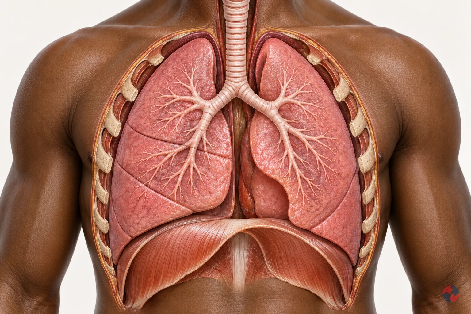

How do the lungs work, and how can workplace exposures affect breathing?
Interactive anatomy · Respiratory tract and lungs · Updated 2026-10-23
Read all anatomy and process explanations without the viewer
Simplified education, not diagnosis, a lung-function or oxygen-saturation test, an exposure calculator or a fitness-to-work assessment. No breath-holding challenge, breathing-rate score or personal health-data collection. Ordinary particle filters do not protect against every gas or oxygen deficiency. COPD and coal-dust topics may reuse normal anatomy only until separate mechanisms are reviewed.
Nose and mouth
Air enters and leaves here. The nose and mouth connect the outside air with the respiratory passages. Air moves through these openings on both the inward and outward breath. Follow the route towards the windpipe before looking at the smaller branches. The illustrated flow is air, a mixture of gases, rather than pure oxygen passing directly into blood. Frimley Health NHS Foundation Trust: How your lungs work · Central and North West London NHS Foundation Trust: How do we breathe?
Trachea
The windpipe carries air into the chest. The trachea, commonly called the windpipe, carries air down towards the chest. It divides into the two main bronchi that supply the lungs. Locate this shared passage before following either branch. Its role is to conduct air towards the gas-exchange regions and provide an outward route for the next exhalation. Frimley Health NHS Foundation Trust: How your lungs work
Bronchi
A main branch carries air into each lung. The two main bronchi take air from the windpipe into the right and left lungs. They divide into progressively smaller branches, distributing the breath through the lungs. Use the split as a landmark before following one highlighted route. These are air passages, distinct from the blood vessels that run through the same organs. Frimley Health NHS Foundation Trust: How your lungs work · University Hospitals of North Midlands NHS Trust: My lung surgery: about your lungs
Bronchioles
Smaller branches distribute air. Bronchioles are smaller branches of the airway system inside the lungs. Their branching routes lead towards clusters of alveoli, where the breath reaches the gas-exchange surface. Follow one branch to its end, using the others as context. This simplified route represents part of a much larger network supplying the lungs together. Frimley Health NHS Foundation Trust: How your lungs work
Alveoli
Tiny air sacs provide a gas-exchange surface. Alveoli are tiny air sacs at the ends of the small airway routes. Their close relationship with surrounding capillaries allows oxygen to pass towards blood and carbon dioxide towards alveolar air. Connect the highlighted cluster with the enlarged gas-exchange view. The enlargement changes the scale so the air space and neighbouring blood vessel can be distinguished. Frimley Health NHS Foundation Trust: How your lungs work · Central and North West London NHS Foundation Trust: How do we breathe?
Air–blood barrier
A thin boundary separates air from blood. The thin alveolar and capillary walls separate air in the sac from blood in its neighbouring vessel. Oxygen and carbon dioxide cross this boundary in opposite directions. Locate the tissue between the two spaces in the enlarged view. Blood cells remain in the vessel; the model does not represent an open connection through which air and blood mix. Central and North West London NHS Foundation Trust: How do we breathe?
Pulmonary capillaries
Small blood vessels pass beside the air sacs. Pulmonary capillaries are small blood vessels beside the alveoli. Blood moving through them receives oxygen and releases carbon dioxide across the thin exchange boundary, then continues through the circulation. Follow the labelled gas routes separately from the blood cells. These cells stay inside the capillary rather than moving into the air sac. Central and North West London NHS Foundation Trust: How do we breathe?
Diaphragm
This muscle helps make room for a breath. The diaphragm is a broad, dome-shaped breathing muscle beneath the lungs. It contracts and flattens downwards during inhalation, helping increase the space in the chest. During a quiet exhalation it relaxes towards its dome shape. Compare it with the rib cage: their changing positions help move air without the lung tissue acting as a contracting muscle. Cambridge University Hospitals NHS Foundation Trust: Breathing techniques to ease breathlessness: muscles involved
Rib cage and breathing muscles
Chest movement changes the space around the lungs. The rib cage protects the chest organs and moves with the breathing muscles. Expansion upwards and outwards helps increase chest space during inhalation; relaxation reduces that space during a quiet exhalation. Compare this movement with the diaphragm below the lungs. Together these structures provide the mechanics that allow air to move through the airways. Central and North West London NHS Foundation Trust: How do we breathe? · Cambridge University Hospitals NHS Foundation Trust: Breathing techniques to ease breathlessness: muscles involved
Pleura
Two thin layers surround the lungs and line the chest. The pleura has two thin layers around the lungs and inside the chest wall. A small amount of fluid between them allows the surfaces to slide with less friction as breathing changes the chest space. Locate this outer covering separately from the internal air sacs; it is not the thin air–blood exchange boundary. Blackpool Teaching Hospitals NHS Foundation Trust: Pleural clinic
Open the complete process transcript
The process, step by step
1 · Make space
Breathing in begins with a change in the space inside the chest. The diaphragm, a dome-shaped muscle beneath the lungs, contracts and moves downwards. Muscles around the ribs help lift the chest upwards and outwards. As the lungs expand with this changing space, air can enter through the airways. Locate the diaphragm and rib cage together rather than treating the lungs as muscles that pull themselves open. The model separates the movement into a teaching step so you can follow its purpose. In the body, chest movement, airflow and blood circulation are connected parts of a continuing process. Central and North West London NHS Foundation Trust: How do we breathe? · Cambridge University Hospitals NHS Foundation Trust: Breathing techniques to ease breathlessness: muscles involved
What to watch: Locate the diaphragm beneath the lungs and the rib cage around them; compare their positions with the space occupied by the lungs.
Why it matters: Changing the chest space draws air into the lung passages. The breathing muscles supply the movement, while the lungs expand with the chest to receive the next breath.
Explore this step: Select the diaphragm and rib cage in turn, then explain how their movement helps air enter without the lungs contracting like muscles.
2 · Move air
Air entering through the nose or mouth travels down the trachea, or windpipe, towards the chest. The trachea divides into the two main bronchi, with one route supplying each lung. Follow this continuous passage in the whole-chest view before moving into the smaller branches. The moving air is a mixture of gases, including oxygen; it is not a stream of pure oxygen travelling straight into the blood. These larger airways carry the breath towards the places where gas exchange happens. The same connected passages also provide the outward route when air is breathed out. Frimley Health NHS Foundation Trust: How your lungs work
What to watch: Start at the nose and mouth, follow the trachea downwards and find the split into the right and left main bronchi.
Why it matters: The larger passages distribute air to both lungs and connect the outside air with the smaller branches inside. Gas exchange takes place further along the route, rather than within the windpipe.
Explore this step: Trace the route to each lung and distinguish the tubes carrying air from the separate vessels carrying blood.
3 · Reach the air sacs
Inside each lung, the bronchi divide into smaller branches called bronchioles. These passages distribute air towards clusters of tiny air sacs, the alveoli. Follow one branch in the close-up and connect its end with an air-sac cluster, using the other branches as surrounding context. These routes distribute the breath throughout the lungs rather than supplying only one working air sac. A real lung contains many branching routes and air sacs operating together. This distribution brings air close to a large network of small blood vessels, preparing for the exchange shown in the next view. Frimley Health NHS Foundation Trust: How your lungs work
What to watch: Follow one smaller airway to the cluster of air sacs at its end, keeping the other branches as surrounding context.
Why it matters: Branching delivers air throughout the lungs instead of to one large chamber. The many air sacs bring the inhaled gases close to blood vessels where oxygen and carbon dioxide can cross.
Explore this step: Trace one complete branch to its air sacs, then explain how the many branches distribute air throughout the lungs.
4 · Change scale
The lesson now changes from a branching airway to an enlarged air–blood interface. An alveolus contains air; beside it runs a capillary containing blood. Their very thin walls form the boundary across which gases can move. Locate the air space, the separating tissue and the inside of the vessel before following any gas symbols. The magnified view represents a tiny part of the same lung, not a separate organ or a hole that joins air and blood into one space. The enlargement helps explain how the two systems can exchange gases while keeping their contents separate. Central and North West London NHS Foundation Trust: How do we breathe?
What to watch: Find the air inside the enlarged alveolus, the thin separating boundary and the neighbouring capillary with blood cells inside it.
Why it matters: A close boundary allows gases to cross between alveolar air and capillary blood. Keeping the two spaces separate lets ventilation and circulation cooperate without blood cells entering the air sac.
Explore this step: Identify the air space and blood-vessel space, then point to the boundary each gas must cross between them.
5 · Exchange gases
At the air–blood boundary, oxygen moves from the alveolar air into the capillary blood. At the same time, carbon dioxide carried back from the body moves from the blood into the air sac. The two gases cross in opposite directions; blood cells stay within the vessel. Follow each labelled symbol separately so that exchanging gases is not confused with mixing the air and blood together. Blood then carries oxygen onwards towards the heart and the body’s tissues. The teaching symbols simplify continuous gas movement, and their size, colour and speed do not represent a measurement. Central and North West London NHS Foundation Trust: How do we breathe?
What to watch: Follow the O₂ symbol towards the capillary and the CO₂ symbol towards the air sac, while locating the blood cells within the vessel.
Why it matters: Oxygen can be carried onwards for use by the body, while carbon dioxide returns to the airways for removal. Both transfers connect the breath with the continuing circulation of blood.
Explore this step: Trace the two gas routes in opposite directions and explain why a blood cell should remain inside the capillary.
6 · Breathe out
During a quiet breath out, the diaphragm and other breathing muscles relax. The diaphragm returns towards its dome shape, chest space decreases and air moves back along the airways to the nose or mouth. This outgoing air carries carbon dioxide that crossed from the blood into the alveoli. Trace the route in reverse without reversing the direction of blood circulation: oxygen-carrying blood continues towards the heart while another breath prepares to enter. The model presents a single cycle to explain the relationship. It does not show breathing as a series of separate events in which circulation stops between steps. Central and North West London NHS Foundation Trust: How do we breathe? · Cambridge University Hospitals NHS Foundation Trust: Breathing techniques to ease breathlessness: muscles involved
What to watch: Compare the diaphragm and chest with the inhalation step, then follow the outward airway route from the lungs to the nose or mouth.
Why it matters: Exhalation removes air containing carbon dioxide from gas exchange. Meanwhile, blood circulation continues carrying oxygen onwards, linking this breathing cycle with the needs of the rest of the body.
Explore this step: Trace the outward air route and the onward blood route separately, then describe why they are connected processes with different pathways.
Read the practice situations and all answer explanations
Sources and how the model is made
The opening images are AI-generated anatomical illustrations. The interactive 3D models and labelled SVG diagrams use original procedural artwork. These teaching images are not medical scans or diagnostic tools. Source references support the learner explanations and do not grant artwork rights.
- Frimley Health NHS Foundation Trust: How your lungs work — Normal airway branching, air sacs and capillary gas exchange; factual text only.
- Central and North West London NHS Foundation Trust: How do we breathe? — Thin alveolar/capillary walls, opposing gas transfer, circulation and chest mechanics. No illustrations or exercises copied.
- Cambridge University Hospitals NHS Foundation Trust: Breathing techniques to ease breathlessness: muscles involved — Diaphragm movement and breathing-muscle context only; this module does not prescribe breathing exercises.
- HSE: Health risks from welding — Welding-related airway irritation, possible deeper injury and irritant asthma; not a universal pattern for every gas.
- HSE: Asthma: an overview — Sensitisation, later triggering and work aggravation. Legacy labelling language and statistics are not reused.
- HSE: Advice for employers: occupational asthma — Exposure prevention, controls and health surveillance only; legacy legislation wording is not reused.
- NHS: Silicosis — Silica-associated inflammation/fibrosis, varying latency, irreversible damage and possible progression after exposure stops.
- HSE: Why is asbestos dangerous? — Asbestosis, mesothelioma and pleural disease are distinct; long disease latency.
- HSE: Construction dust: cutting paving blocks, kerbs and flags — Task design, source suppression/extraction, suitable RPE and checking controls; not a generic dust safe-dose rule.
- NHS: Shortness of breath — Emergency recognition of severe breathing difficulty and 999; emergency advice is outside the activity.
- NHS: Asthma — Narrowed/swollen airways, symptoms and clinical diagnosis; no medication doses or treatment plan reproduced.
- University Hospitals of North Midlands NHS Trust: My lung surgery: about your lungs — Right/left lung lobe counts and airway anatomy; no source image copied.
- Blackpool Teaching Hospitals NHS Foundation Trust: Pleural clinic — Pleural layers and the small lubricating fluid layer outside lung tissue; no procedural teaching included.
- HSE: Asphyxiation hazards in welding and allied processes — Oxygen-deficient spaces and need for independent air supply; no unprotected entry or rescue.
- HSE: Respiratory protective equipment at work — RPE selection must match the contaminant and conditions; filtering protection is not a supply of oxygen.
- HSE: Flour dust and risks to bakers — Flour sensitisation and practical reduction of dusty handling; exposure-limit numbers are not reused.
- Health and Safety Executive: Asbestos essentials — Task planning, training and competent asbestos work arrangements for the maintenance exposure example; not permission to disturb unknown material or a licence/classification decision.
- Health and Safety Executive: Welding fume: protect your workers — Alternative joining, fume reduction, LEV, suitable RPE and maintained controls for the welding-fume example. No unprotected exposure or rescue procedure.
What is the exposure-to-action pathway?
The practical pathway is source, inhalation, reaction, symptoms, exposure removal, control investigation and health review.
This topic becomes much easier to understand when it is followed as a chain. A contaminated source is disturbed; fine organic dust, spores or aerosol enter the breathing zone; a susceptible person inhales the material; the immune system reacts in the lungs; symptoms appear after the task or shift; the exposure is stopped or reduced; controls are investigated; and occupational health or medical advice is used where symptoms suggest possible work-related disease. HSENI occupational lung disease
The pathway also helps managers avoid two common mistakes: treating symptoms only as ordinary sickness, and treating dust only as a housekeeping problem. The question is whether work is repeatedly creating an inhalable biological or organic exposure.
| Step | What happens | Workplace decision |
|---|---|---|
| Exposure source | Mouldy hay, grain, compost, leaf mould, bird material, wood dust or contaminated ventilation material is present. | Identify and record the source before disturbing it. HSENI occupational lung disease |
| Inhaled organic dust or aerosol | Fine material becomes airborne and reaches the breathing zone. | Stop dry disturbance, ventilate, segregate and control the task. HSENI occupational lung disease Water-system source |
| Immune reaction | A susceptible person may develop lung inflammation after exposure. | Do not diagnose locally; recognise the possible work link. HSENI occupational lung disease |
| Symptoms | Fever, cough, breathlessness and fatigue may appear after exposure and improve away from work. | Record the task pattern and signpost health advice. HSENI occupational lung disease |
| Remove exposure | The worker should not continue the same uncontrolled exposure while the issue is investigated. | Pause or redesign work until controls are verified. HSENI occupational lung disease |
| Investigate controls | COSHH, cleaning, ventilation, RPE, supervision and reporting arrangements are checked. | Update assessment, briefings and evidence. HSENI occupational lung disease |
| OH/medical review | Qualified health input considers symptoms and work history. | Use work-focused advice while protecting confidentiality. HSENI occupational lung disease |
Do not confuse hypersensitivity pneumonitis with other respiratory issues
Different respiratory diseases can overlap in symptoms, so the page must help readers separate the workplace control questions.
Cough and breathlessness are not enough to identify the disease. Occupational asthma, COPD, humidifier fever, infection and ordinary dust irritation can all be confused with hypersensitivity pneumonitis. That is why occupational history, exposure pattern, medical assessment and competent risk assessment are important. HSENI occupational lung disease
The control message remains practical: investigate the source, reduce exposure, use official health routes and avoid sending people back into the same uncontrolled conditions.
| Issue | Typical workplace clue | Do not confuse with | Workplace action |
|---|---|---|---|
| Hypersensitivity pneumonitis | Symptoms after organic dust, mould, bird, animal or contaminated aerosol exposure. | Ordinary cold or simple nuisance dust. | Review COSHH controls and use OH/medical advice. HSENI occupational lung disease |
| Occupational asthma | Wheeze, chest tightness or variable symptoms linked to sensitiser exposure. | Farmer's lung or COPD without assessment. | Use asthma risk assessment, exposure control and health surveillance. HSENI occupational lung disease |
| Humidifier fever | Flu-like symptoms linked to contaminated mist or water systems. | Legionnaires' disease or farmer's lung. | Isolate source, investigate water/mist controls and signpost health advice. HSE HSG132 sick building syndrome System controls |
| COPD | Longer-term breathing limitation that may be affected by dusts, fumes or smoking history. | Acute feverish episodes only. | Control dust/fume exposure and support health assessment. HSENI occupational lung disease |
Where can exposure arise?
Exposure can arise anywhere organic or biological material becomes airborne and reaches the breathing zone.
Mouldy hay, straw, grain and stored vegetable matter are documented farmer’s-lung routes. Compost, roof voids, humidifiers and wood dust can pose other bioaerosol or respiratory hazards, but this page does not label every such exposure a proven HP cause; assess the agent and clinical evidence separately. Grain fever is also distinct from farmer’s lung. HSE work-related respiratory disease statistics HSE grain dust
Traditional examples include a farmer handling mouldy hay, straw, grain or stored crops in a poorly ventilated barn. Modern workplaces may also see risks in estate teams clearing damp compost or leaf mould, woodworking shops handling fine organic dust, facilities teams opening contaminated ventilation sections or humidification systems, animal bedding, bird-contaminated roof voids, mushroom cultivation, contaminated building voids and mist-producing systems.
The higher-risk pattern is dry, disturbed, poorly ventilated, enclosed or repeated work where fine dust or aerosol is generated close to the worker.
| Source | Work example | Why it matters | Control focus |
|---|---|---|---|
| Mouldy hay, straw or grain | Handling stored crop material in barns or silos. | Spores and microbial material can become airborne. | Avoid mouldy material where possible, ventilate, control dust by assessed enclosed or mechanised methods and use suitable RPE where needed. HSE grain dust |
| Compost and green waste | Turning compost, clearing vegetation or handling mouldy leaves. | Potential bioaerosol or respiratory hazard; HP attribution needs agent-specific exposure and clinical evidence. | Mechanise, segregate, control dust by assessed enclosed or mechanised methods and avoid dry dusty disturbance. HSE agriculture COSHH task guidance |
| Bird or animal material | Roof void clearance, nesting material, animal bedding or droppings. | Potential bioaerosol or respiratory hazard; HP attribution needs agent-specific exposure and clinical evidence. | Survey, isolate, clean safely and use competent controls. HSE agriculture COSHH task guidance |
| Woodworking organic dust | Sanding, sweeping or machining wood in poorly controlled workshops. | Potential bioaerosol or respiratory hazard; HP attribution needs agent-specific exposure and clinical evidence. | Use LEV, housekeeping, RPE where needed and health reporting. HSE woodworking wood dust |
| Contaminated aerosol | Misting, humidification, poorly maintained water-based systems. | Potential water-system aerosol hazard; HP attribution needs agent-specific exposure and clinical evidence. | Maintain systems, clean reservoirs and stop suspect sources. HSE Legionella workplace risk systems |
| Enclosed dusty work | Poorly ventilated stores, lofts, plant rooms or barns. | Exposure concentration rises quickly. | Ventilate, restrict access and plan work methods. HSE grain dust |
What symptoms and patterns should trigger action?
Symptoms should be interpreted by clinicians, but work patterns should be noticed by managers and supervisors.
Possible patterns include feverish episodes, chills, cough, chest tightness, breathlessness, fatigue, weight loss or symptoms that worsen after a particular task, shift or area and improve away from work exposure. Repeated reports from several people doing similar work are a strong warning sign. HSE grain dust
People may not connect symptoms with work at first. Briefings should tell workers to report recurring breathing symptoms and to mention the tasks they were doing.
| Pattern | Why it matters | Action |
|---|---|---|
| Flu-like illness after dusty work | May be dismissed as seasonal illness. | Record task and exposure details and review controls. HSE grain dust |
| Breathlessness or chest tightness | May indicate significant respiratory effect. | Remove from exposure and signpost to health advice. HSENI occupational lung disease |
| Several workers affected | Could indicate shared exposure source. | Escalate as a cluster and investigate source. HSENI occupational lung disease |
| Symptoms improve away from work | Possible work-related pattern. | Use occupational health and exposure review. HSENI occupational lung disease |
| Symptoms recur after return | Controls may still be inadequate. | Do not re-expose without competent review. HSENI occupational lung disease |
How should the risk be assessed?
Assessment should connect the source, airborne route, exposed people, controls and health follow-up.
A suitable assessment should observe the work. It should not rely only on a material name or a generic dust assessment. Ask what is being disturbed, whether it is mouldy, dry or contaminated, how it becomes airborne, who is in the breathing zone, how long the task lasts and whether symptoms have been reported. HSENI occupational lung disease
The assessment should also check cleaning methods. Dry sweeping, compressed air and uncontrolled disturbance can make exposure worse.
| Question | Why it matters | Evidence |
|---|---|---|
| What organic or biological source is present? | Identifies the likely antigen or contaminated material. | Task survey, photos, material description and worker input. HSENI occupational lung disease |
| How does it become airborne? | Controls must target inhalation. | Observation, dust/aerosol description and ventilation review. HSENI occupational lung disease |
| Who is exposed? | Bystanders, cleaners and contractors may be missed. | Exposure group map and work schedule. HSENI occupational lung disease |
| What symptoms have been reported? | Early warning signs may already exist. | Confidential symptom route and OH referral trigger. HSENI occupational lung disease |
| Are controls verified? | Paper controls may not work in dusty enclosed tasks. | Supervisor checks, RPE fit testing and maintenance records. HSENI occupational lung disease |
How can the hierarchy of controls reduce organic-dust exposure and Farmer's Lung risk?
Controls should follow ERICPD and prioritise source control before relying on respiratory protection.
Verify controls: Handling mould-affected hay


Read the complete visual as text
- Actual task: A farm finds mould-affected hay. Avoid unnecessary disturbance and plan stock, animal-health and disposal decisions with competent advice.
- Evidence to check: stock condition, planned handling/disposal, dust control and selected RPE
- Stop or hold condition: uncontrolled mould dust, failed cab/filter control or respiratory symptoms requiring review
- Release evidence and owner: farm manager accepts the controlled handling route and effective exposure protection
Verify controls: Cleaning an empty grain store


Read the complete visual as text
- Actual task: A worker removes settled grain dust from an empty accessible store; assess organic dust, machinery, fire and access hazards separately.
- Evidence to check: store access, machinery isolation, dust-safe collection and task-suitable RPE
- Stop or hold condition: uncontrolled dust cloud, ignition risk, failed isolation or an unplanned confined-space requirement
- Release evidence and owner: store owner accepts clean conditions and repaired dust controls
Prevent dust generation and use engineering controls before relying on RPE. Do not dry sweep or use compressed air on grain dust. For necessary residual exposure, choose suitable RPE with fit testing and supervision; generic gloves or aprons do not control inhalation. HSE grain dust HSE respiratory protective equipment
The best control is to avoid generating the exposure. Where work must continue, reduce dust or aerosol at source, ventilate, enclose or segregate work, clean without raising dust and use suitable respiratory protective equipment only where residual risk remains.
RPE must be suitable, face-fit tested where tight-fitting, maintained and worn correctly. It should not become a substitute for avoiding mouldy, dry, dusty work methods.
Choose the activity
How can the ERICPD hierarchy of controls reduce risks while handling mould-affected hay?

| Control level | Control for this activity | Owner and verification | Stop / restart decision |
|---|---|---|---|
| Eliminate | Reject mould-affected feed for use and avoid unnecessary disturbance. Plan safe removal of the affected stock; outsourcing removal does not eliminate the collector’s dust exposure. | Process owner verifies that the revised workflow achieves the required output and the hazardous step is genuinely removed. | If the hazardous step remains, retain a task assessment and select the stronger feasible remaining controls. |
| Reduce, including substitution | Use suitable sound stock or a validated lower-dust feed form after checking animal and process requirements. | Process/technical owner validates the alternative, its compatibility and any new risks before adoption. | Reject the alternative if suitability is unproved or its new hazards are unresolved. |
| Isolate | Segregate affected stock and keep other workers and occupied areas away from the disturbance and dust path. | Supervisor verifies the task boundaries and that the separation or isolation protects affected people and connected work. | Hold or stop for uncontrolled mould dust, failed cab/filter control or respiratory symptoms requiring review. |
| Control: physical safeguards | Use suitable mechanised handling, enclosed filtered cabs and effective dust controls; avoid shaking or breaking material by hand. | Competent equipment/process owner checks stock condition, planned handling/disposal, dust control and selected RPE. | Hold or stop for uncontrolled mould dust, failed cab/filter control or respiratory symptoms requiring review. |
| Control: work method and response | Plan the minimum necessary disturbance, weather, hygiene and disposal; arrange appropriate health surveillance and symptom reporting. | Supervisor confirms the limits, response, communication and restart authority with the people doing this activity. | Stop if the agreed method, communication, competent cover or task limits cannot be maintained. |
| PPE | Select adequate suitable RPE for agricultural dust and mould exposure, with fit testing for tight-fitting facepieces; nuisance masks are unsuitable. | Assessor confirms whether PPE is needed and checks selection, condition, compatibility and any required face-fit evidence. | Do not rely on unsuitable or defective protection, or use PPE to continue after an essential source control fails. |
| Discipline | Check dust controls, cab filters and stock condition and review storage causes before new supplies are accepted. | Release only after farm manager accepts the controlled handling route and effective exposure protection. Retain the checks and unresolved restrictions at handover. | Keep the activity on hold while defects or required reinstatement evidence remain unresolved. |
How can the ISO 45001 hierarchy of controls reduce risks while handling mould-affected hay?

| Control level | Control for this activity | Owner and verification | Stop / restart decision |
|---|---|---|---|
| Elimination | Reject mould-affected feed for use and avoid unnecessary disturbance. Plan safe removal of the affected stock; outsourcing removal does not eliminate the collector’s dust exposure. | Process owner verifies that the revised workflow achieves the required output and the hazardous step is genuinely removed. | If the hazardous step remains, retain a task assessment and select the stronger feasible remaining controls. |
| Substitution | Use suitable sound stock or a validated lower-dust feed form after checking animal and process requirements. | Process/technical owner validates the alternative, its compatibility and any new risks before adoption. | Reject the alternative if suitability is unproved or its new hazards are unresolved. |
| Engineering controls | Segregate affected stock and keep other workers and occupied areas away from the disturbance and dust path. | Supervisor verifies the task boundaries and that the separation or isolation protects affected people and connected work. | Hold or stop for uncontrolled mould dust, failed cab/filter control or respiratory symptoms requiring review. |
| Engineering controls | Use suitable mechanised handling, enclosed filtered cabs and effective dust controls; avoid shaking or breaking material by hand. | Competent equipment/process owner checks stock condition, planned handling/disposal, dust control and selected RPE. | Hold or stop for uncontrolled mould dust, failed cab/filter control or respiratory symptoms requiring review. |
| Administrative controls | Plan the minimum necessary disturbance, weather, hygiene and disposal; arrange appropriate health surveillance and symptom reporting. | Supervisor confirms the limits, response, communication and restart authority with the people doing this activity. | Stop if the agreed method, communication, competent cover or task limits cannot be maintained. |
| PPE | Select adequate suitable RPE for agricultural dust and mould exposure, with fit testing for tight-fitting facepieces; nuisance masks are unsuitable. | Assessor confirms whether PPE is needed and checks selection, condition, compatibility and any required face-fit evidence. | Do not rely on unsuitable or defective protection, or use PPE to continue after an essential source control fails. |
| Administrative controls | Check dust controls, cab filters and stock condition and review storage causes before new supplies are accepted. | Release only after farm manager accepts the controlled handling route and effective exposure protection. Retain the checks and unresolved restrictions at handover. | Keep the activity on hold while defects or required reinstatement evidence remain unresolved. |
How can the ERICPD hierarchy of controls reduce risks while cleaning an empty grain store?

| Control level | Control for this activity | Owner and verification | Stop / restart decision |
|---|---|---|---|
| Eliminate | Use a suitable enclosed storage design that can be cleaned externally where feasible, removing routine occupied entry for dust removal. Existing stores still need an assessed cleaning method. | Store designer confirms that inspection, dust removal and foreseeable blockage clearance can occur externally; record any entry that the design still requires. | If the hazardous step remains, retain a task assessment and select the stronger feasible remaining controls. |
| Reduce, including substitution | Use a suitable mechanised collection or dust-controlled cleaning method instead of hand dry-brushing. | Process/technical owner validates the alternative, its compatibility and any new risks before adoption. | Reject the alternative if suitability is unproved or its new hazards are unresolved. |
| Isolate | Securely isolate grain-handling machinery and separate the cleaning zone from occupied work and vehicle routes. | Supervisor verifies the task boundaries and that the separation or isolation protects affected people and connected work. | Hold or stop for uncontrolled dust cloud, ignition risk, failed isolation or an unplanned confined-space requirement. |
| Control: physical safeguards | Use suitable dust-safe vacuum collection and extraction; do not improvise equipment for combustible dust or blow deposits into the air. | Competent equipment/process owner checks store access, machinery isolation, dust-safe collection and task-suitable RPE. | Hold or stop for uncontrolled dust cloud, ignition risk, failed isolation or an unplanned confined-space requirement. |
| Control: work method and response | Plan access, waste and cleaning products. Silo/confined-space entry needs a separate assessed method and rescue arrangements. | Supervisor confirms the limits, response, communication and restart authority with the people doing this activity. | Stop if the agreed method, communication, competent cover or task limits cannot be maintained. |
| PPE | For the small-scale HSE AG9 cleaning route, provide at least APF 20 RPE and any additional product protection; fit-test tight-fitting facepieces. | Assessor confirms whether PPE is needed and checks selection, condition, compatibility and any required face-fit evidence. | Do not rely on unsuitable or defective protection, or use PPE to continue after an essential source control fails. |
| Discipline | Check the cleaned store, equipment and ventilation and verify that dust-control defects are repaired before refilling. | Release only after store owner accepts clean conditions and repaired dust controls. Retain the checks and unresolved restrictions at handover. | Keep the activity on hold while defects or required reinstatement evidence remain unresolved. |
How can the ISO 45001 hierarchy of controls reduce risks while cleaning an empty grain store?

| Control level | Control for this activity | Owner and verification | Stop / restart decision |
|---|---|---|---|
| Elimination | Use a suitable enclosed storage design that can be cleaned externally where feasible, removing routine occupied entry for dust removal. Existing stores still need an assessed cleaning method. | Store designer confirms that inspection, dust removal and foreseeable blockage clearance can occur externally; record any entry that the design still requires. | If the hazardous step remains, retain a task assessment and select the stronger feasible remaining controls. |
| Engineering controls | Use a suitable mechanised collection or dust-controlled cleaning method instead of hand dry-brushing. | Process/technical owner validates the alternative, its compatibility and any new risks before adoption. | Reject the alternative if suitability is unproved or its new hazards are unresolved. |
| Engineering controls | Securely isolate grain-handling machinery and separate the cleaning zone from occupied work and vehicle routes. | Supervisor verifies the task boundaries and that the separation or isolation protects affected people and connected work. | Hold or stop for uncontrolled dust cloud, ignition risk, failed isolation or an unplanned confined-space requirement. |
| Engineering controls | Use suitable dust-safe vacuum collection and extraction; do not improvise equipment for combustible dust or blow deposits into the air. | Competent equipment/process owner checks store access, machinery isolation, dust-safe collection and task-suitable RPE. | Hold or stop for uncontrolled dust cloud, ignition risk, failed isolation or an unplanned confined-space requirement. |
| Administrative controls | Plan access, waste and cleaning products. Silo/confined-space entry needs a separate assessed method and rescue arrangements. | Supervisor confirms the limits, response, communication and restart authority with the people doing this activity. | Stop if the agreed method, communication, competent cover or task limits cannot be maintained. |
| PPE | For the small-scale HSE AG9 cleaning route, provide at least APF 20 RPE and any additional product protection; fit-test tight-fitting facepieces. | Assessor confirms whether PPE is needed and checks selection, condition, compatibility and any required face-fit evidence. | Do not rely on unsuitable or defective protection, or use PPE to continue after an essential source control fails. |
| Administrative controls | Check the cleaned store, equipment and ventilation and verify that dust-control defects are repaired before refilling. | Release only after store owner accepts clean conditions and repaired dust controls. Retain the checks and unresolved restrictions at handover. | Keep the activity on hold while defects or required reinstatement evidence remain unresolved. |
When may health surveillance or occupational health be needed?
Repeated or credible respiratory exposure should be managed with competent occupational health input where the risk justifies it.
HSE says health surveillance is appropriate when a disease or adverse effect is associated with the exposure, a valid way exists to detect it early and reduce harm, and workplace conditions make it likely. Repeated exposure alone is insufficient. Clinical records remain confidential; managers receive work-focused conclusions. HSE COSHH health surveillance ICO workers health information
Health surveillance is not a generic annual form. It should be linked to the exposure, the possible disease and a valid way to detect early harm. For respiratory risks, this may include symptom questionnaires, lung function testing and referral routes where concerns arise.
Health records and medical records are different. Managers need fitness, restrictions and control-learning information, not unnecessary diagnosis detail.
| Trigger | What to consider | Evidence |
|---|---|---|
| Repeated exposure to organic dust | Whether a respiratory health surveillance scheme is needed. | OH risk advice and scheme scope. HSE grain dust |
| Reported symptoms | Prompt referral and exposure review. | Work-focused referral and action tracker. HSENI occupational lung disease |
| Cluster of symptoms | Possible uncontrolled source. | Incident review, source investigation and worker communication. HSENI occupational lung disease |
| New task or process | Whether controls and surveillance need updating. | COSHH review and training record. HSENI occupational lung disease |
| Return after symptoms | Whether further exposure is safe. | OH advice and control verification. HSENI occupational lung disease |
What evidence shows the risk is controlled?
Evidence should show that exposure sources are known, controls are used and early warning signs are acted on.
Records should prove that controls were selected, implemented, checked and improved. The table helps distinguish useful evidence from records that exist only because someone expected a document. COSHH Regulations 2002
| Record | Purpose | Review trigger |
|---|---|---|
| COSHH assessment | Links source, route, exposed people, controls and health follow-up. | New task, symptoms or control failure. COSHH Regulations 2002 |
| Organic dust register | Identifies recurring sources such as mouldy crop, compost, bird material or contaminated aerosol. | New location, season or contractor task. HSE grain dust |
| RPE records | Shows selection, fit testing, inspection and maintenance. | Face change, new RPE or poor use. HSE respiratory protective equipment |
| Health surveillance record | Tracks scheme participation without unnecessary medical detail. | Symptom trend or exposure change. HSE COSHH health surveillance |
| Cleaning and waste plan | Prevents dry sweeping and uncontrolled disturbance. | Audit finding or complaint. HSENI occupational lung disease |
| Action tracker | Shows defects and control improvements are closed. | Overdue action or repeated symptoms. HSENI occupational lung disease |
How can this appear in a real workplace?
Illustrative stored-crop task
For mouldy hay/grain, first decide whether disturbance is necessary. Assess storage, transfer and airborne release; keep bystanders out and verify the selected enclosure/capture or handling method before starting. Select residual RPE for the actual task and wearer, distinguishing fit testing from each-use checks. New or worsening breathing symptoms need clinical referral now, regardless of whether a surveillance programme is established. Do not deliberately re-expose someone to test a work/rest pattern; return depends on clinical advice and verified controls. The roof-void case remains a separate suspected bioaerosol example, not a proved HP diagnosis.
A scenario helps people remember the exposure route and control decisions.
Worked workplace scenario — part 1 of 2


Read the complete visual as text
- Discovery: Weak response: Carry on and clean quickly. Better response: Stop, isolate area and assess contamination. Evidence: Stop-work note and task review.
- Method: Weak response: Dry sweep and bag waste. Better response: Use controlled cleaning and avoid raising dust. Evidence: Method statement and supervisor check.
- People: Weak response: Assume only cleaners are exposed. Better response: Identify workers, contractors and nearby occupants. Evidence: Exposure group list.
Worked workplace scenario — part 2 of 2


Read the complete visual as text
- Health: Weak response: Treat symptoms as ordinary sickness. Better response: Signpost to health advice and review work link. Evidence: OH referral route and action log.
- Learning: Weak response: Do the same next time. Better response: Add roof void and organic dust prompt to surveys. Evidence: Updated checklist and briefing.
A leisure site opens a closed roof void before refurbishment. Workers find old nesting material, dry organic dust and a musty smell. The first instinct is to sweep it out quickly before the contractor arrives, but one worker reports feverish symptoms and a tight chest after similar work the previous year. HSENI occupational lung disease
A stronger response is to stop, assess the material, restrict access, plan controlled removal, avoid dry sweeping, use competent cleaning methods, brief workers on symptoms and check whether occupational health advice is needed.
| Decision point | Weak response | Better response | Evidence |
|---|---|---|---|
| Discovery | Carry on and clean quickly. | Stop, isolate area and assess contamination. | Stop-work note and task review. HSENI occupational lung disease |
| Method | Dry sweep and bag waste. | Use controlled cleaning and avoid raising dust. | Method statement and supervisor check. HSENI occupational lung disease |
| People | Assume only cleaners are exposed. | Identify workers, contractors and nearby occupants. | Exposure group list. HSENI occupational lung disease |
| Health | Treat symptoms as ordinary sickness. | Signpost to health advice and review work link. | OH referral route and action log. HSENI occupational lung disease |
| Learning | Do the same next time. | Add roof void and organic dust prompt to surveys. | Updated checklist and briefing. HSENI occupational lung disease |
Which duties apply in Great Britain and Northern Ireland?
Use separate GB and NI disease-reporting decisions
Northern Ireland RIDDOR 1997 regulation 5 and Schedule 3 include extrinsic alveolitis (including farmer’s lung) with specified work/exposure conditions. The responsible reporting person checks the medical diagnosis and actual work link against that route. In GB, distinguish the regulation 8 named list from any applicable regulation 9 occupational biological-agent route; neither automatically includes nor excludes every HP case. Reporting does not delay individual clinical assessment.
NI RIDDOR regulation 5: occupational disease · NI RIDDOR Schedule 3 · HSE: Occupational carcinogens, mutagens and biological agents
Choose the route for the actual site, task and trigger; guidance and clinical advice do not replace nation-specific law.
COSHH is the primary hazardous biological dust route. HP is not automatically a named GB RIDDOR disease; check exact specified criteria if a diagnosed case raises reporting questions. COSHH Regulations 2002 COSHH Regulations (NI) 2003 HSE reportable occupational diseases
| Decision | Great Britain law | Northern Ireland law | Applicability |
|---|---|---|---|
| General health and safety | Health and Safety at Work etc. Act 1974 GB law | Health and Safety at Work (NI) Order 1978 NI law | Work activity |
| Risk assessment and arrangements | Management of Health and Safety at Work Regulations 1999 GB law | Management of Health and Safety at Work Regulations (NI) 2000 NI law | Organic-dust task |
| Hazardous biological dust | COSHH Regulations 2002 GB law | COSHH Regulations (NI) 2003 NI law | Credible exposure to hazardous biological material |
How should the selected controls be checked in practice?
ERICPD is used here as a planning mnemonic. The linked HSE five-level hierarchy is the official source for the order and relative reliability of controls; the two labels are not identical.
Start with source removal or work design, then engineering and administrative controls; use PPE only for residual exposure.
ERICPD is a planning mnemonic: Isolate generally maps to engineering, Discipline sustains controls, and the five-level hierarchy names substitution separately. Each control requires assessment of the actual task. HSE five-level hierarchy HSE five-level hierarchy
How should management verify controls for this task?
For mouldy stored-crop dust inhalation and suspected farmer’s lung, the management check should start with a real control decision: Dispose of badly mouldy stock using a planned method. Name the person who will test it, keep evidence from the actual work, and reopen the assessment when the failure condition in the hierarchy table occurs. HSE management-system guidance.
A review should compare the planned control with what workers and supervisors can use in practice. Record the defect, owner, interim protection, verification and next review trigger so that a policy statement or certificate does not stand in for a working control.
How could Riverside Leisure Ltd manage this in practice?

Riverside Leisure Ltd would manage hypersensitivity pneumonitis risk through exposure recognition, controlled cleaning, worker reporting and occupational-health escalation.
Riverside Leisure Ltd is a leisure and holiday park business with grounds teams, maintenance staff, roof voids, plant rooms, composting areas, lakeside buildings and occasional animal or bird contamination. Its risk is not farming in the traditional sense, but many of the same organic dust and bioaerosol lessons apply. HSENI occupational lung disease
| Management element | How the business would manage it | Evidence to review |
|---|---|---|
| Assessing | Riverside adds mouldy organic material, nesting material, compost and contaminated aerosol sources to its COSHH and premises inspection prompts. | Organic dust source register and COSHH review. HSENI occupational lung disease |
| Analysing | HSE and facilities review tasks where dust is dry, enclosed, repeated or reported with symptoms. | Task observation and symptom trend review. HSENI occupational lung disease |
| Implementing | Dry sweeping is prohibited for suspect material; controlled removal, ventilation, exclusion and suitable RPE are specified. | Method statement, RPE records and supervisor sign-off. HSENI occupational lung disease |
| Training | Grounds, cleaning and maintenance teams are briefed on symptom reporting and stop-work triggers. | Toolbox talk and induction records. HSENI occupational lung disease |
| Checking | First-line supervisors inspect work areas; second-line audits sample COSHH records and RPE evidence. | Inspection report and action tracker. HSENI occupational lung disease |
| Learning | Any respiratory symptom cluster after a task triggers exposure review and management review. | Incident learning and revised survey checklist. HSENI occupational lung disease |
What must each role demonstrate?
Record an observed decision, assessor, evidence and trigger for reassessment.
Attendance alone does not prove that a worker or manager can recognise the hold, referral or review point. HSENI occupational lung disease
| Role | Demonstration | Assessor and record | Reassess when |
|---|---|---|---|
| Worker | Recognise dusty source and recurring breathlessness. | Supervisor observes stop/report. | New material or symptoms. HSENI occupational lung disease |
| Farm lead | Assess mouldy stored-crop dust and stop unsafe work. | Competent assessor verifies. | Storage/process changes. HSE grain dust |
| Controls engineer | Check ventilation and dust capture. | Independent test record. | Control failure. HSE agriculture COSHH task guidance |
| RPE assessor | Select and fit-test RPE for residual dust. | Fit-test record. | Facepiece or wearer changes. HSE respiratory protective equipment |
| OH lead | Assess surveillance criteria and confidential referral. | Clinical governance review. | Symptoms or exposure changes. HSE COSHH health surveillance |
What warning signs show controls are weak?
Weak controls usually show up as dusty work, recurring symptoms, poor cleaning methods or missing evidence.
Warning signs are included because failure usually appears before a serious event. Treat these points as prompts for inspection, audit, supervision and management review rather than as simple compliance statements. HSE grain dust
| Warning sign | How harm can develop | Action |
|---|---|---|
| Dry sweeping organic dust | Fine material can be inhaled deeply. | Stop and use controlled cleaning. HSE grain dust |
| Musty enclosed area | Mould or microbial material may be present. | Assess before disturbance. HSENI occupational lung disease |
| Repeated flu-like symptoms | Work-related respiratory disease may be missed. | Escalate to OH and exposure review. HSENI occupational lung disease |
| No RPE fit testing | Respiratory protection may not work. | Review selection, fit test and training. HSE respiratory protective equipment |
| Symptoms treated as absence only | Exposure source remains uncontrolled. | Use incident and health surveillance routes. HSENI occupational lung disease |
Chemical hazard symbols: what do they mean?
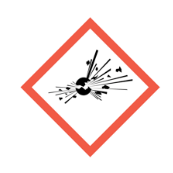
Exploding bomb
Explosive, self-reactive or organic peroxide hazards.
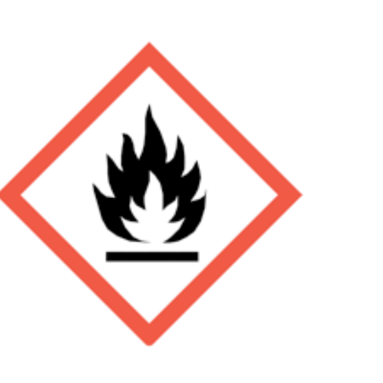
Flame
Flammable, self-heating or pyrophoric substances.
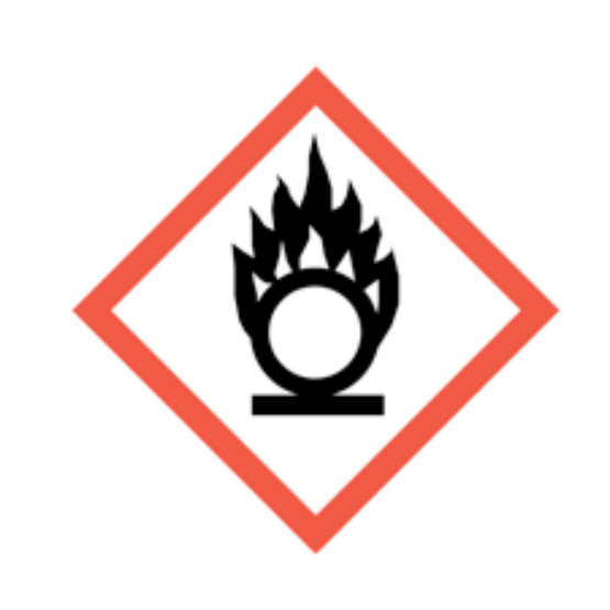
Flame over circle
Oxidising substances that can intensify fire.

Gas cylinder
Gas under pressure.
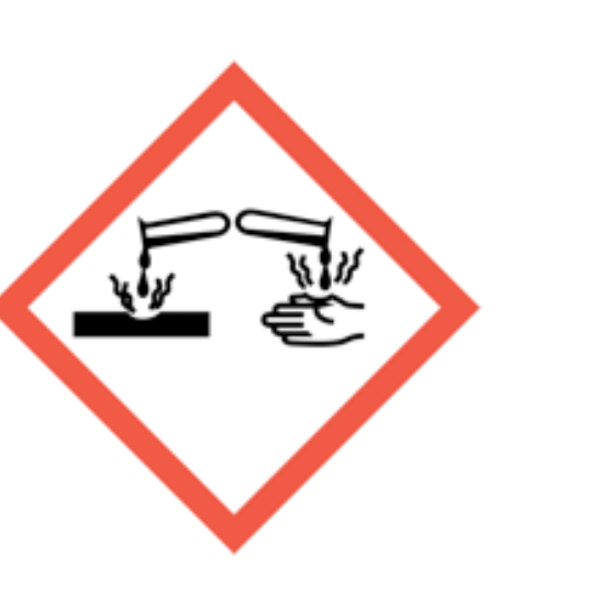
Corrosion
Skin burns, eye damage or corrosive to metals.
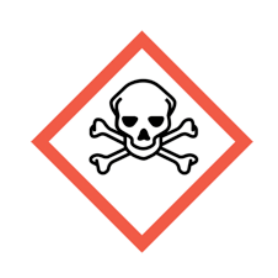
Skull and crossbones
Acute toxicity.

Exclamation mark
Irritation, harmful effects or skin sensitisation; some classifications cover drowsiness, dizziness or ozone-layer hazard. Read the actual hazard statements. Skin sensitisation differs from respiratory sensitisation.
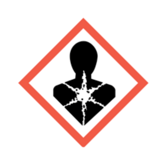
Health hazard
Serious health hazards, including organ damage, aspiration hazard, respiratory sensitisation, cancer and reproductive effects; some can follow a single exposure.
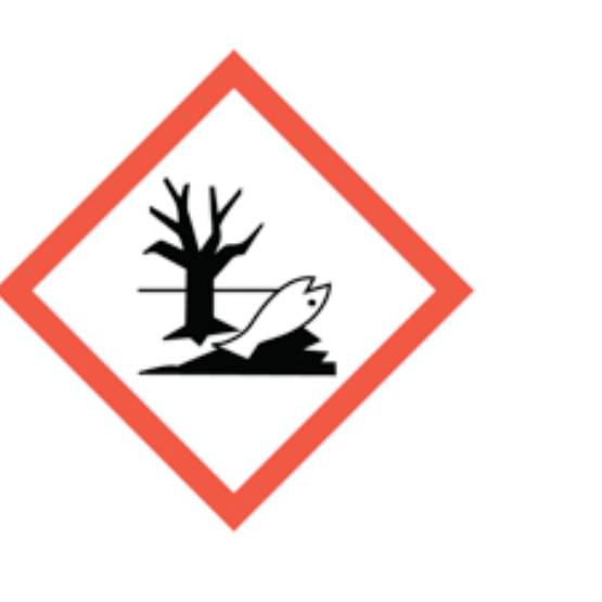
Environment
Hazardous to the aquatic environment.
What do the acronyms and key terms mean?
Acronyms should be understood before they are used in assessments, briefings, audits or learning answers.
Acronyms can exclude newer readers if they are not explained. This section keeps the main terms available at the end of the page so the article stays readable while still supporting learners and audit users.
| Acronym | Meaning | Plain-English use |
|---|---|---|
| COSHH | Control of Substances Hazardous to Health | UK regulations for hazardous substances, including biological agents where relevant. |
| EAA | Extrinsic allergic alveolitis | Another term often used for hypersensitivity pneumonitis. |
| OH | Occupational health | Qualified work-focused health advice. |
| RPE | Respiratory Protective Equipment | Breathing protection used where exposure remains after better controls. |
| LEV | Local Exhaust Ventilation | Engineering control that captures airborne contaminants at source. |
Unlock another resource
You have used this month’s two free downloads. Publish a post in your own words to unlock one more resource, or choose SHARE Individual for 10 downloads per month.
You decide whether to share. If you do, use the relevant platform disclosure tools and do not make claims you do not genuinely hold.